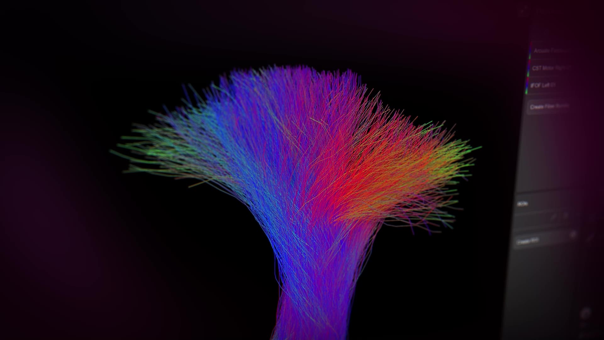
elements-fibertracking-mp4

Elements FibertrackingMatter that matters

Discover more

Brainlab Elements Fibertracking revolutionizes neurosurgery with the constrained spherical deconvolution (CSD) analysis module.1 This software enables the visualization and analysis of brain white matter pathways for enhanced neurological understanding and patient care.

Switching between deterministic diffusion tensor imaging (DTI)-based and probabilistic CSD-based tractography enables users to focus on critical and patient-specific visualization.

1_1informyourdecisions_4k-1-scaled.jpg
2_v2_2usetrackingtemplates_af2_4k-1-scaled.jpg
3_3unlockplanning_4k-scaled.jpg

Inform your decisions with advanced tracking1

Take full advantage of research-grade probabilistic tractography in your clinical practice to identify multiple fiber directions like crossing and fanning fibers, as well as fibers that may be present in edema.

I believe having multiple algorithms or models within the same planning software is beneficial. It allows us to view brain structures from more rigid to more flexible perspectives, enabling fine-tuning between models to achieve the most accurate anatomical outcomes. Moreover, the introduction of probabilistic tractography is one of the most significant advancements to reconstruct the complexity of white matter architecture.

Francesco Latini, MD, PhD, University Hospital Uppsala, Uppsala, Sweden
elements_background-scaled.jpg

Pathways of significance


CSD*-based
tractography visualizes fiber tracts more accurately than DTI

offering potentially improved safety margin estimates crucial for neurosurgical planning.


Source: Farquharson S et al. (2013): J Neurosurg., DOI: 10.3171/2013.2.JNS121294


*Constrained spherical deconvolution


Up to %
of brain voxels exhibit multiple fiber orientations

highlighting the necessity of employing a higher order tractography model like CSD, known for its superior ability to detect crossing fibers.

Source: Jeurissen B et al. (2013): Hum Brain Mapp., DOI: 10.1002/hbm.22099; Tournier JD et al. (2008): Neuroimage, DOI: 10.1016/j.neuroimage.2008.05.002


Improved tracking
through edema

may help clinicians achieving a safe maximal
resection.

Source: Yeh FC et al. (2021): Neuroimage, DOI: 10.1016/j.neuroimage.2021.118651

Elevate your neurosurgical approach. Explore state-of-the-art solutions for surgery.

240314_elements_surgery_1445x380px.png

One suite for all your planning needs

Brainlab Elements interactive and semi-automated planning modules for state-of-the-art planning for cranial image guided surgery.

More information to elevate your decision-making

In addition to deterministic fiber tracking, Elements Fibertracking gives you the option to see even more with the addition of probabilistic fiber tractography. Switch between these two modes to visualize complex fiber tracts near tumor or edema regions.1

4-1_v2_dti-tractography_cst_4k-scaled.jpg

DTI-based deterministic tractography

Deterministic DTI-based fiber tracking provides a robust single and dual tensor model able to reconstruct straightforward pathways, aiding visualization and interpretation of results.

CSD-based probabilistic tractography1

Probabilistic CSD-based fiber tracking is an advanced tracking method that allows for the identification of multiple fiber directions within a voxel, overcoming the limitation of the DTI-based technique when dealing with crossing, fanning or high angulation fibers, or when navigating through regions of edema.

Use cases for fiber tracking

Elements Fibertracking provides you with the information you need to make your informed decisions.

Cranial resection procedures

For cranial resection procedures, such as eloquent and non-eloquent tumors, skull-base tumors, or other lesions, the avoidance of critical structures is crucial. Elements Fibertracking may support in these critical procedures by visualizing cranial white matter.

5_usecaseelotumorresection_anonymous.jpg

Functional neurosurgery

During functional neurosurgery procedures, white matter tracts can serve either as target regions or areas to avoid. Therefore, tractography information can inform DBS trajectory planning or be correlated with implanted sEEG electrodes and support subsequent epilepsy resection or ablation strategies.

image-2024-06-12-17_34_06.png

Your roadmap to the brain starts here

Experience state-of-the-art solutions for surgery.

Related products

240314_elements_surgery_relatedproduct.jpg

Cranial Planning

Our Brainlab Elements software modules are designed to assist clinical teams in state-of-the-art planning for image guided cranial surgery.

Cranial Microscope Integration with Navigation Software

Microscope Navigation

Microscope Navigation injects relevant data directly into the ocular, providing surgeons meaningful context without distracting from the surgical situation.

Screenshot: Cranial Navigation Application

Cranial Navigation

Our Cranial Navigation system combines ease of use with extended functionality, supporting informed approach planning and confident navigation, tailored to meet surgeon needs during cranial procedures.

  1. Not yet commercially available. Please contact your sales representative.