Screenshot: Cranial Navigation Application

Cranial Navigation ApplicationDetailed visualization.
Intuitive control.

Brainlab neuronavigation combines ease of use with extended functionality tailored to surgeons’ needs

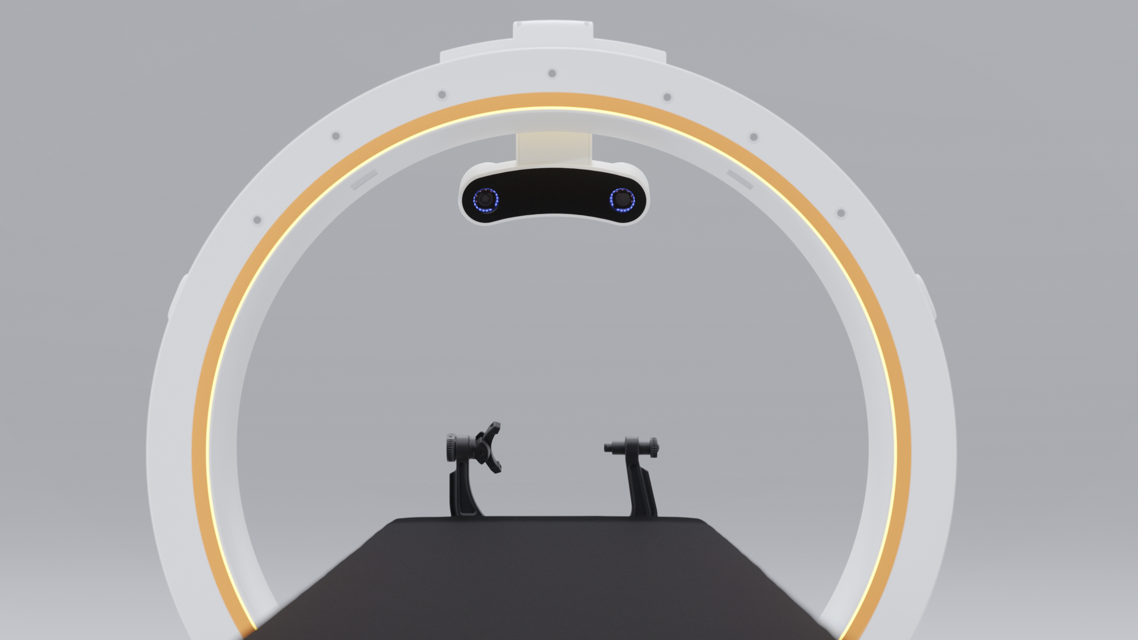
Automatic Registration with Loop-X

Intraoperative X-Ray images acquired by Loop-X can be seamlessly transferred to Brainlab Navigation. Regardless of the patient’s position, especially for cases in prone position, Automatic Registration with Loop-X simplifies the registration process and automates the workflow. Loop-X allows patient positioning within its 121 cm gantry opening and supports non-isocentric image acquisition for patient-tailored O.R. setup.

  • Scalable volume between 3 x 3 cm and 25 x 48 cm

  • Low dose scan protocol available

  • Dynamic Collimation during 2D and 3D scans helps protect sensitive anatomical structures

  • Loop-X may support intraoperative visualization of cranial structures such as ventricles and detect lesions and intraoperative bleeding (ICB)

  • Intraoperative implant verification in cranioplasty cases

  • Intraoperative imaging for pituitary cases and skull base procedures

20240319_loopx_update_rendering_08_00006.jpg

Automatic Registration for iMRI, iCT and other imaging modalities

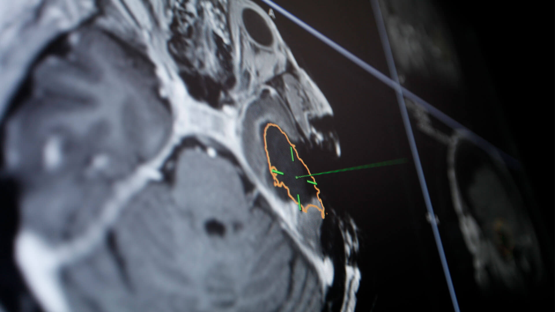
Automatic Image Registration is a fundamental technology for seamless integration of intraoperative MRI (iMRI) and other imaging devices with image-guided surgery. This technology adds intraoperatively acquired data to surgical navigation, optimizing the workflow while maintaining the preoperative surgical plan.

  • Accurate, automatic image registration without the need to access anatomical landmarks 

  • Automatic Registration for iMRI realizes the automatic image registration via markers integrated in head holder1

  • AIR iMRI available for various MR scannersUniversal Automatic Registration technology available for integration of other imaging devices such as iCT, rotational angiograph and cone-beam CT1

Automatic Image Registration (AIR) technology in action

Surface Matching Registration

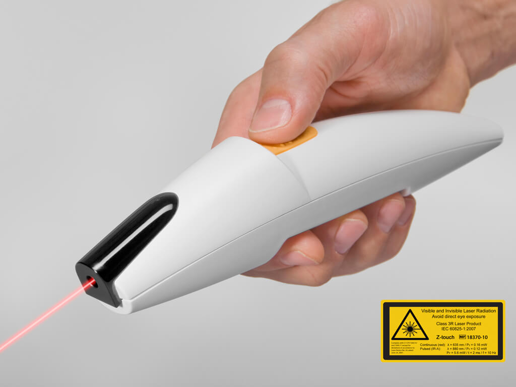
Z-touch® and Softouch® are time-saving2 registration methods based on surface matching, making patient registration easy by simply touching the patient’s skin with either a pointer or a laser. As both devices register existing diagnostic CT and MR images without headsets or markers, additional registration scans can be avoided, helping to reduce neuronavigation costs and radiation exposure to the patient and O.R. team.

  • Pointer registration with or without markers

  • Touch-sensitive surface matching with Softouch

  • Contact-free surface matching with Z-touch

  • Combined surface matching registration

  • Pre- and intraoperative landmark registration

Photo of Z-touch® for Time-Saving Registration in Brainlab Cranial Navigation

Approach Planning

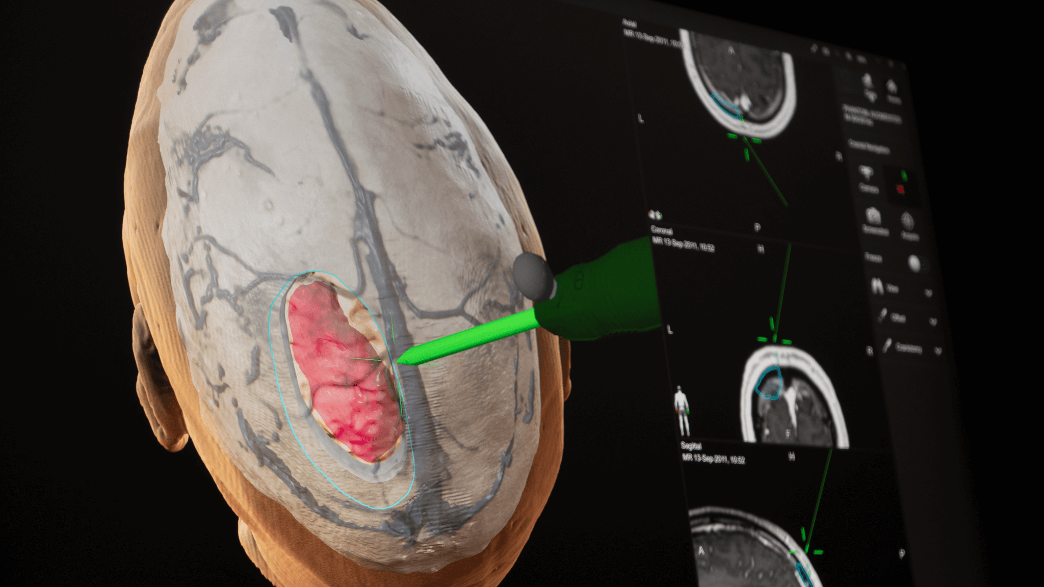
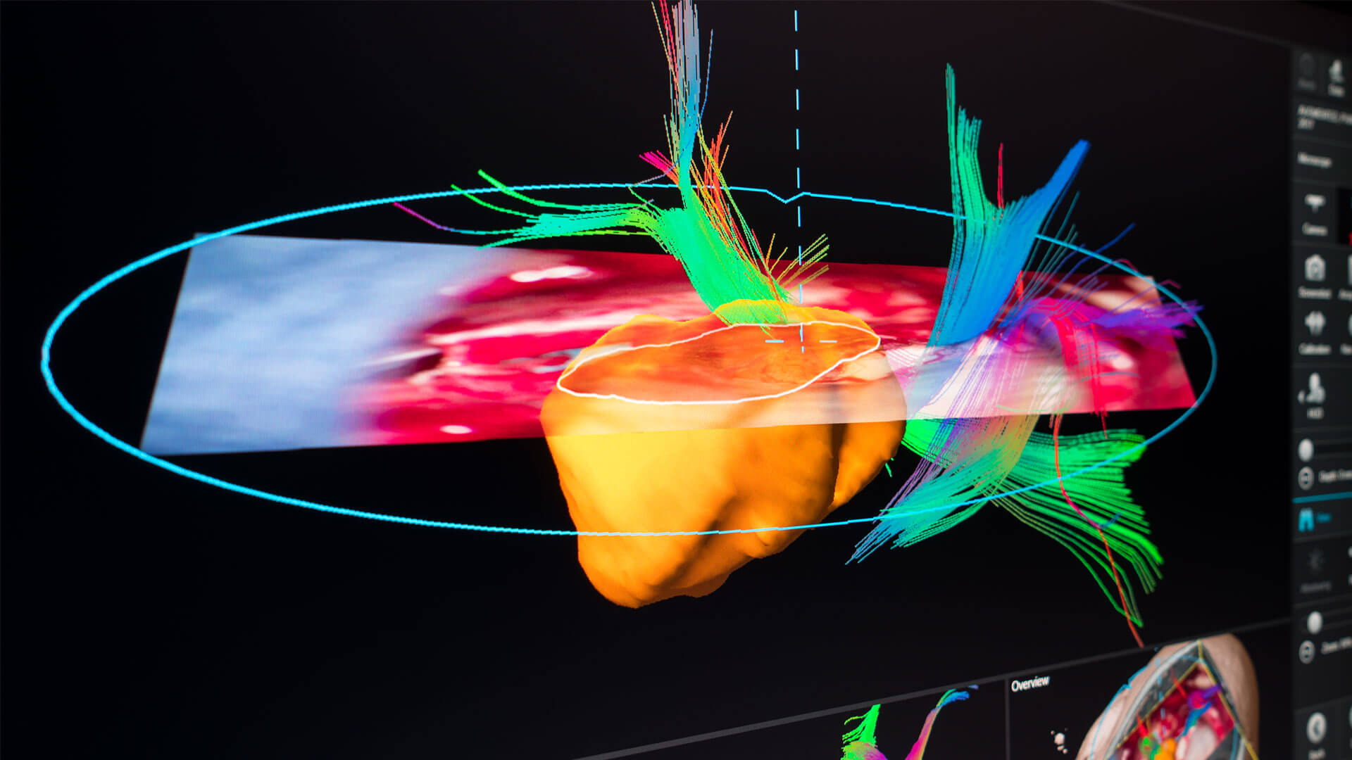
Informed approach planning requires consolidated clinical data. Cranial Navigation summarizes relevant and detailed anatomical information in one dedicated view helping surgeons identify the optimal craniotomy before making the incision.

  • Automatic segmentation of skin, bone, cortical vessels and cerebrum

  • 3D preview of the lesion and critical structures before opening the skull

  • Virtual scalpel simulates craniotomy and creates a bone flap object that can be saved for documentation

  • Approach planning in 3D and correlated inline or axial, coronal, sagittal navigation views provides spatial orientation

  • Dedicated Maximum Intensity Projection (MIP)-based visualization of surface vessels contributes to more informed patient access planning

Screenshot: Cranial Navigation - Virtual scalpel simulating craniotomy

Easy-to-use Navigation

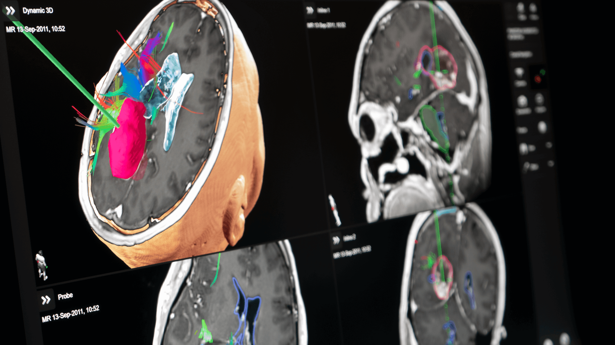
When navigating, distraction-free concentration is essential. Detailed visualizations are tailored to show only clinically relevant data allowing the surgeon to focus on what’s necessary for their work. Automated data selection and layout composition are designed to reduce screen interaction, making Cranial Navigation even more user-friendly.3

  • Smart Approach view dynamically adapts and zooms according to resection progress

  • Overlay mode with fused CT and ultrasound data consolidates anatomical details

  • Dynamic 3D view provides 3D context around the tool tip to help with orientation

  • User interface automatically displays patient data in the most suitable layout

Screenshot: Detailed visualizations in Cranial Navigation

Vascular Navigation

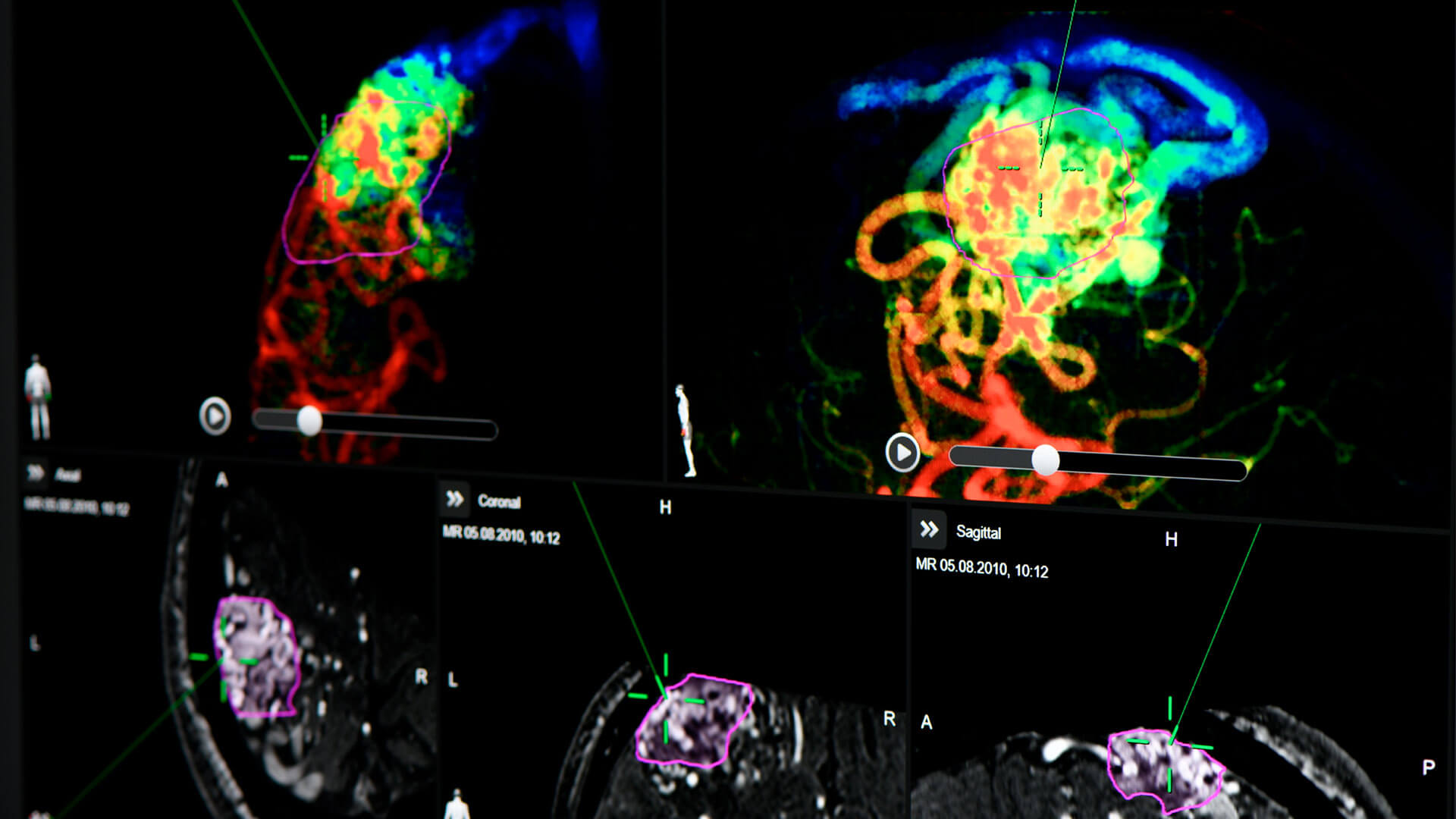
Cranial Navigation Angio4 helps surgeons to identify feeding and draining veins on navigated digital subtraction angiography (DSA) data or locate a nidus object with dedicated view layouts. Co-registration of angiographies and subsequent neurovascular contouring adds clinical context from anatomical 3D data contributing to improved orientation.3

  • Combines co-registered angiography pairs and navigated MR/CT reconstructions

  • Interactively displays dynamic color-coded contrast flow projections to assist with surgical target identification and analysis

  • Visualizes 2D DSA data as Color Intensity Projection (CIP) to help identify feeding and draining vessels

  • Displays nidus object on navigation data

Screenshot: Cranial Navigation Angio - ubsequent neurovascular contouring

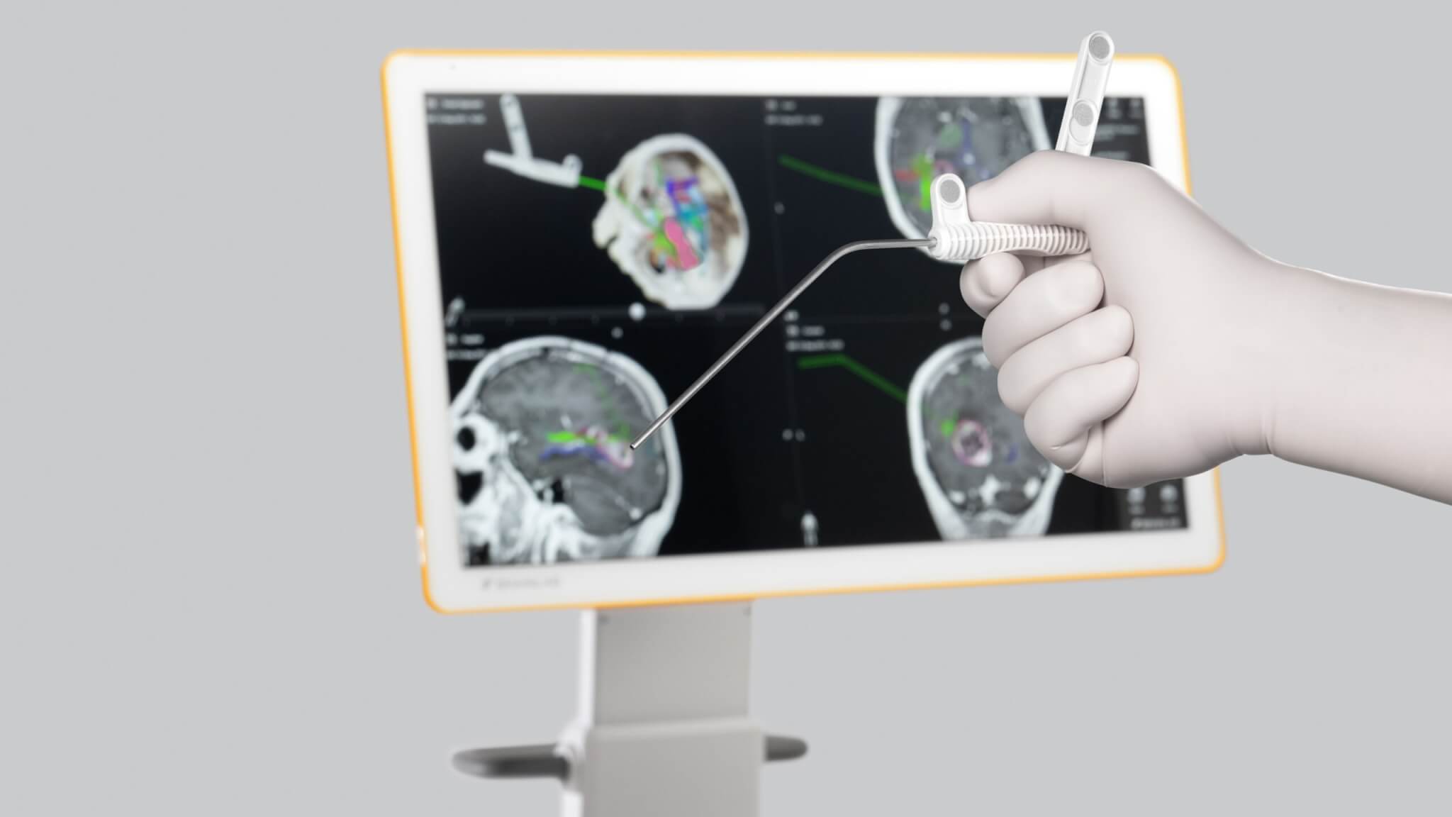
Navigated Disposable Stylet

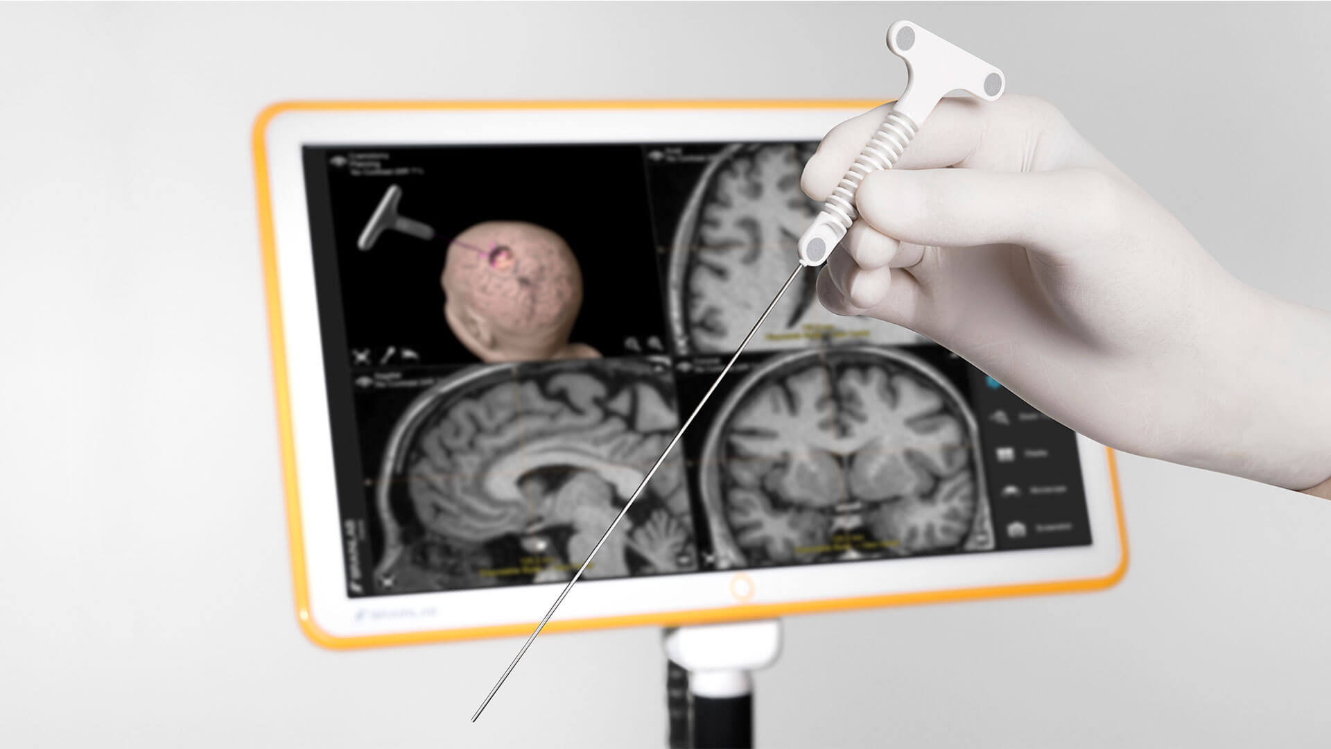
The Disposable Stylet is designed for the navigated placement of shunts or ventricular catheters in neurosurgery, enabling their freehand placement. The incorporated passive marker geometry is automatically recognized by the navigation camera, allowing for instant use.

  • Navigation-ready pre-calibrated tool

  • Shunt/catheter vendor independent

  • 1.1 mm diameter, 165 mm length

Navigated Disposable Stylet

Robotic Biopsy Alignment

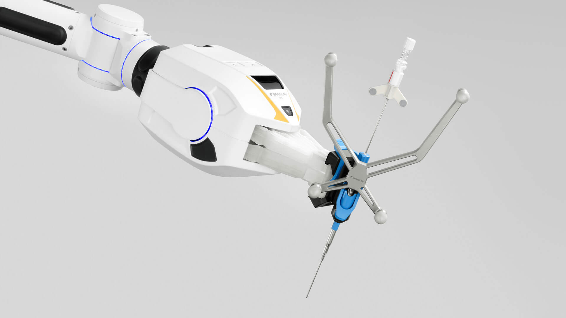
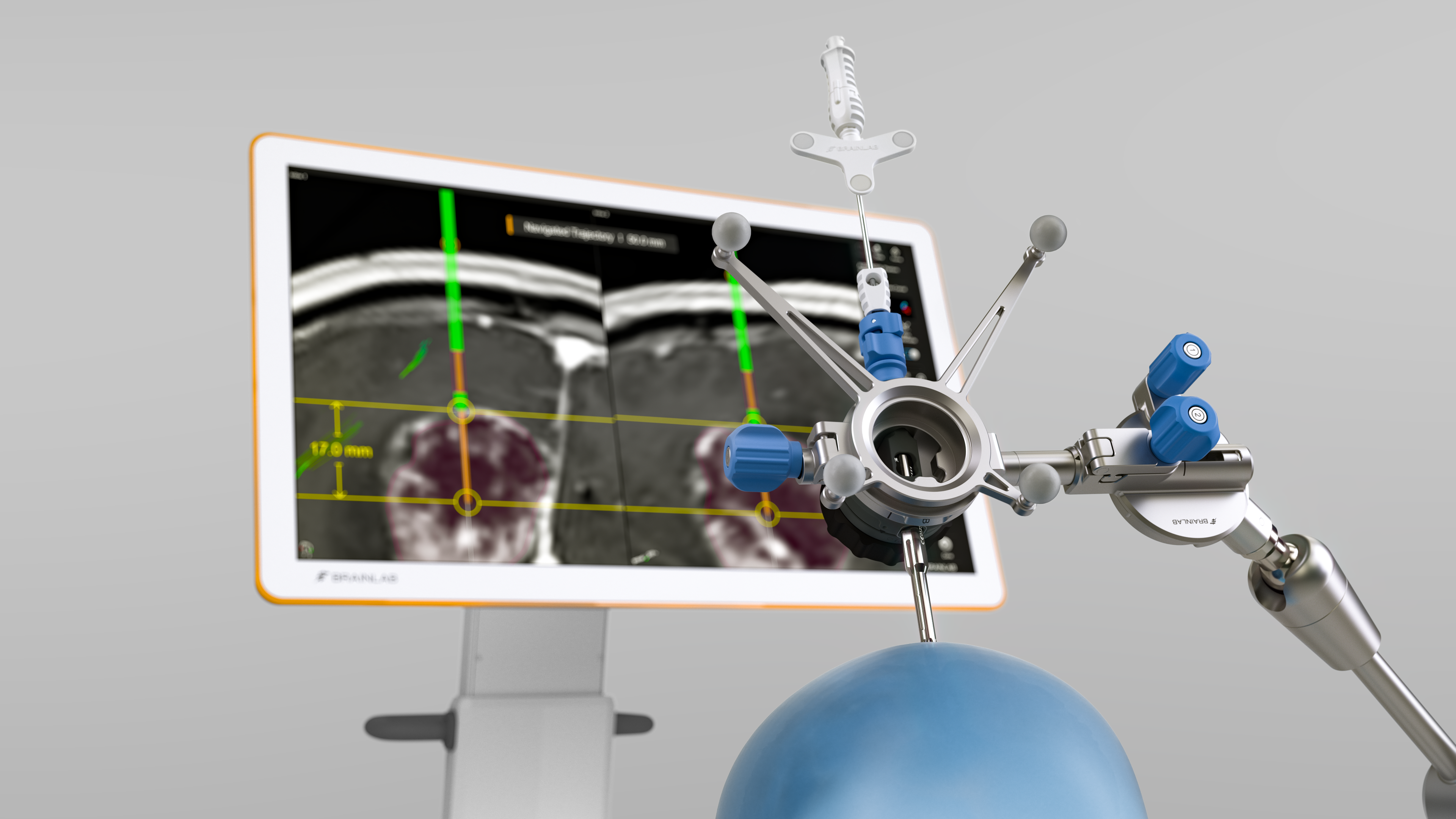
The Cirq alignment software facilitates intuitive manual arm positioning while the robotic module performs automatic trajectory alignment. Trajectories can conveniently be defined using Elements Trajectory Planning or the on-the-fly instrument offset acquisition within Cranial Navigation.

  • Automatic alignment to planned biopsy trajectory after manual arm positioning

  • Cirq alignment software provides visual guidance towards region of interest

  • Biopsy anchor for the skull adds stability to the setup

Photo of Cirq: Robotic Biopsy Alignment

VarioGuide Alignment

Surgeons can target pre-planned trajectories with Brainlab VarioGuide® Alignment System. The frameless instrument holder combined with the VarioGuide Alignment Software Cranial achieve precision and increase efficiency and patient comfort during navigated frameless biopsies.5

  • Frame-based precision without a head frame5

  • Flexibly adapts to instruments from Ø 1.8 – 8 mm

  • Dedicated (optional) Cranial Biopsy Drill Kit minimizes the potential for deviation from the planned trajectory and allows for minimally invasive drilling

  • Software-supported measurement of bone thickness for setting the Drill Depth Stop prevents drilling too deep

  • Potential reduction of total procedure time5

  • Disposable Pre-calibrated Biopsy Needle (Ø 2.1 mm) seamlessly integrates into navigation workflow to reach planned trajectories

ns_img_varioguidebiopsyneedle2-0_oct24_rev3.png

Navigated Disposable Suction

The Disposable Pre-calibrated Suction6 combines the functionality of a classic suction and a navigated pointer into one unique instrument. Its ergonomic, lightweight design supports seamless navigation, especially under the microscope.

  • Pre-calibrated, ready-to-navigate suction and pointer in one tool

  • Integrated passive markers on both sides for right and left-handed use

  • Available with outer tube diameter of 2.7 mm (Charr 8) and 4.0 mm (Charr 12)

  • Lightweight design (14 g or 18 g) for comfortable instrument handling

  • Materials do not contain natural rubber latex, therefore suitable for surgeries on patients with latex allergies

Disposable Pre-calibrated Suction

Augmented Reality Visualizations

Augmented reality has become an essential technology in neurosurgery, virtually showing surgeons what lies beneath the surface. Microscope Navigation digitally overlays the planned surgical target and surrounding structures as semi-transparent objects onto the real patient anatomy. These visualizations provide anatomical insight and spatial orientation throughout the procedure.

  • Provides meaningful context about the surgical situation without having to look away from it

  • Maintains navigation accuracy and corrects initial patient registration based on anatomical landmarks

  • Navigation-controlled robotic movement supports ergonomic and accurate alignment to navigated instruments and anatomical landmarks

  • Seamlessly integrates with major surgical microscope vendors

  • Auto-detects connected microscopes, minimizing setup effort3

Augmented Reality Visualization through Microscope Navigation

Live Intraoperative Imaging

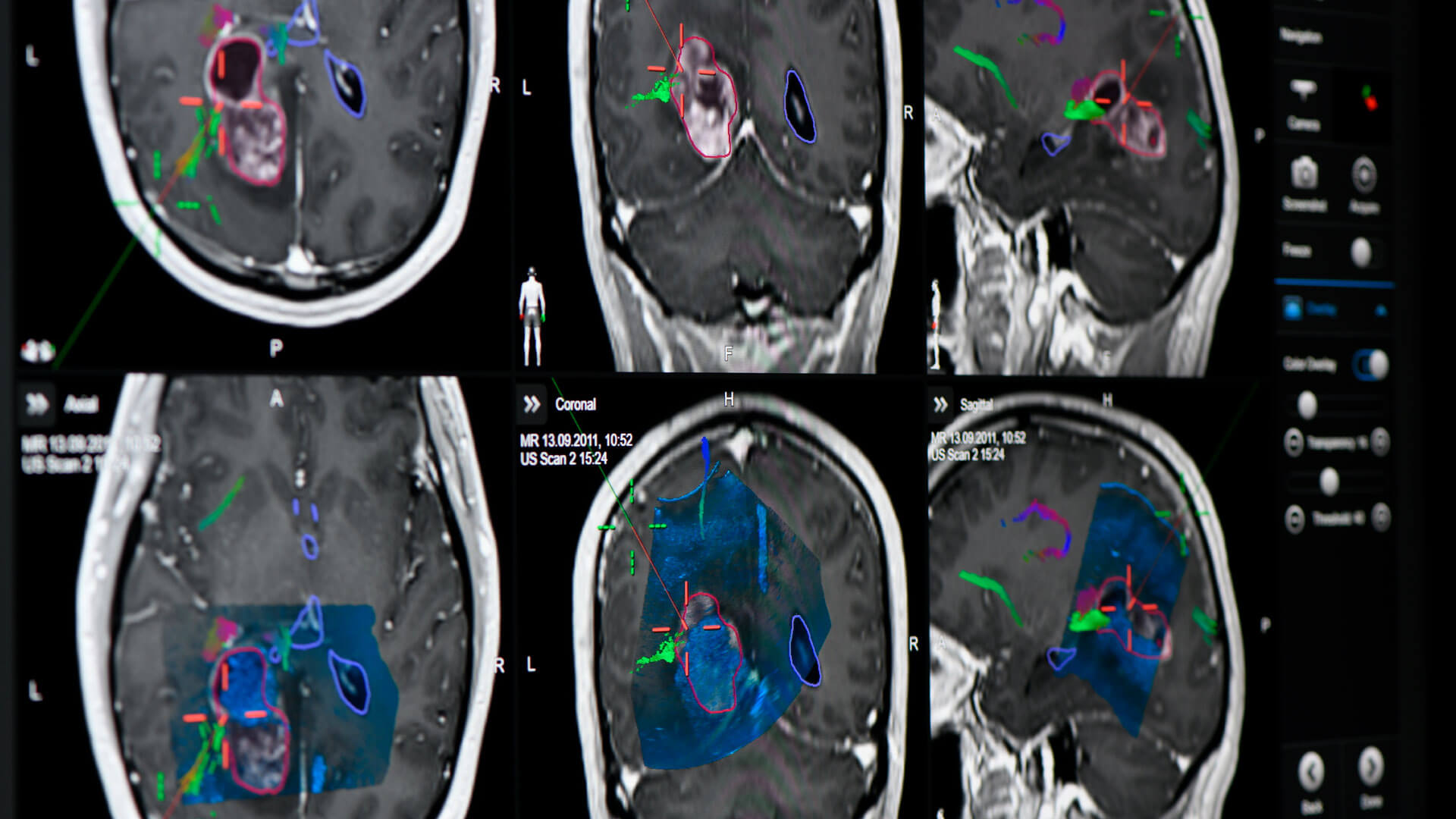
Ultrasound Navigation provides real-time intraoperative imaging that complements cranial image guidance. With a small footprint and minimal interruption of the surgical workflow compared to other imaging modalities, it’s a suitable addition for almost every neurosurgical suite.

  • Overlays real-time ultrasound images in 2D or 3D

  • Provides accurate orientation as well as updates on resection progress

  • Allows for visualization and assessment of brain shift

  • 3D scanning of the cavity helps identify residual tumor and helps achieve the planned extent of resection

Screenshot of the Ultrasound Navigation: real-time intraoperative imaging for cranial image guidance

Intraoperative Neuromonitoring

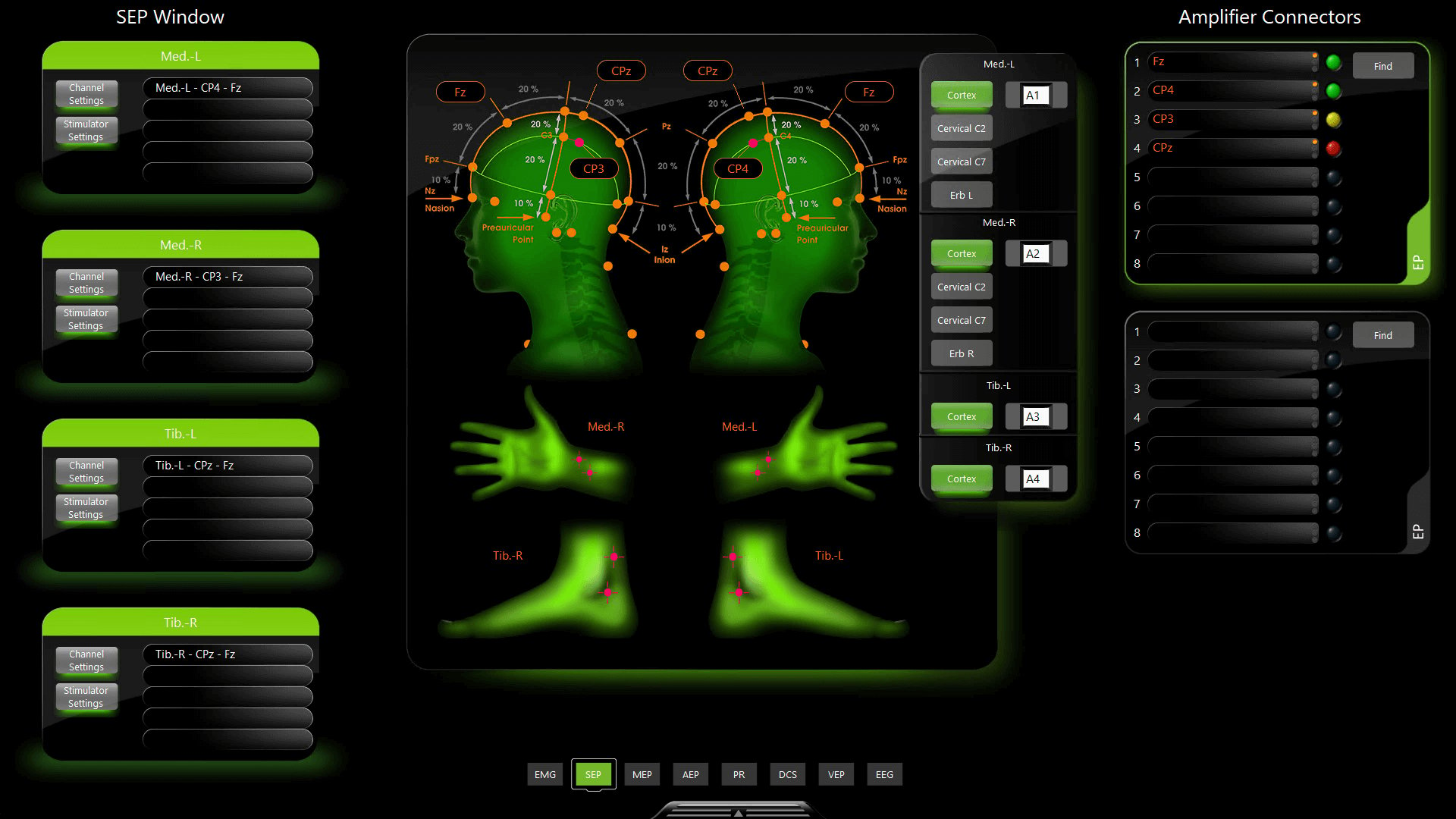
Intraoperative neuromonitoring (IONM) supports decision making during surgery with live functional information about the nervous system. The Langer Medical neuromonitoring system AVALANCHE® Plus7 combines powerful monitoring and mapping functionalities with intuitive setup and ease of use.

  • Enables verification and refinement of preoperative planning data from Fibertracking, using mapping techniques

  • Streamlines the setup process: select among available configurations and customize them through anatomy-guided touch screen interaction

  • Ensures consistent signal quality by visualizing the electrode impedances according to a traffic light system

  • Facilitates the creation of a surgical report and postoperative analysis through event marking and screenshot functionalities

Screenshot: Langer Medical neuromonitoring system AVALANCHE® Plus

Technical Information

Flyer VarioGuide Alignment System Cranial Biopsy

Flyer Disposable Stylet

Flyer Disposable Suction

  1. Technical feasibility determined on case-by-case basis

  2. Compared to extra time required for MR or CT scan to perform fiducial-based registration

  3. Compared to previous version: Cranial/ENT

  4. Requires Elements Planning Application

  5. Bradac et al. (2017): World Neurosurgery, DOI:10.1016/j.wneu.2017.04.104

  6. Not yet commercially available in several countries (FDA clearance available). Please contact your sales representative.

  7. Not yet commercially available in several countries. Please contact your sales representative.