Brainlab Elements Fibertracking révolutionne la neurochirurgie avec le module d’analyse de déconvolution sphérique contrainte (CSD)1. Ce logiciel permet la visualisation et l’analyse des voies de la substance blanche du cerveau pour une meilleure compréhension neurologique et des soins aux patients.
Le passage d’une tractographie déterministe basée sur l’imagerie du tenseur de diffusion (DTI) à une tractographie probabiliste basée sur la CSD permet aux utilisateurs de se concentrer sur la visualisation critique et spécifique au patient.


Découvrez notre technologie grâce aux experts cliniques
Je crois que disposer de plusieurs algorithmes ou modèles dans le même logiciel de planification est bénéfique. Il nous permet de visualiser les structures cérébrales d’une perspective plus rigide à une perspective plus souple, ce qui permet un réglage fin entre les modèles pour obtenir les résultats anatomiques les plus précis. En outre, l’introduction de la tractographie probabiliste est l’une des avancées les plus significatives pour reconstruire la complexité de l’architecture de la substance blanche.

Voies de communication importantes
Améliorez votre approche neurochirurgicale. Explorez de solutions chirurgicales de pointe.

Plus d’informations pour améliorer votre prise de décision
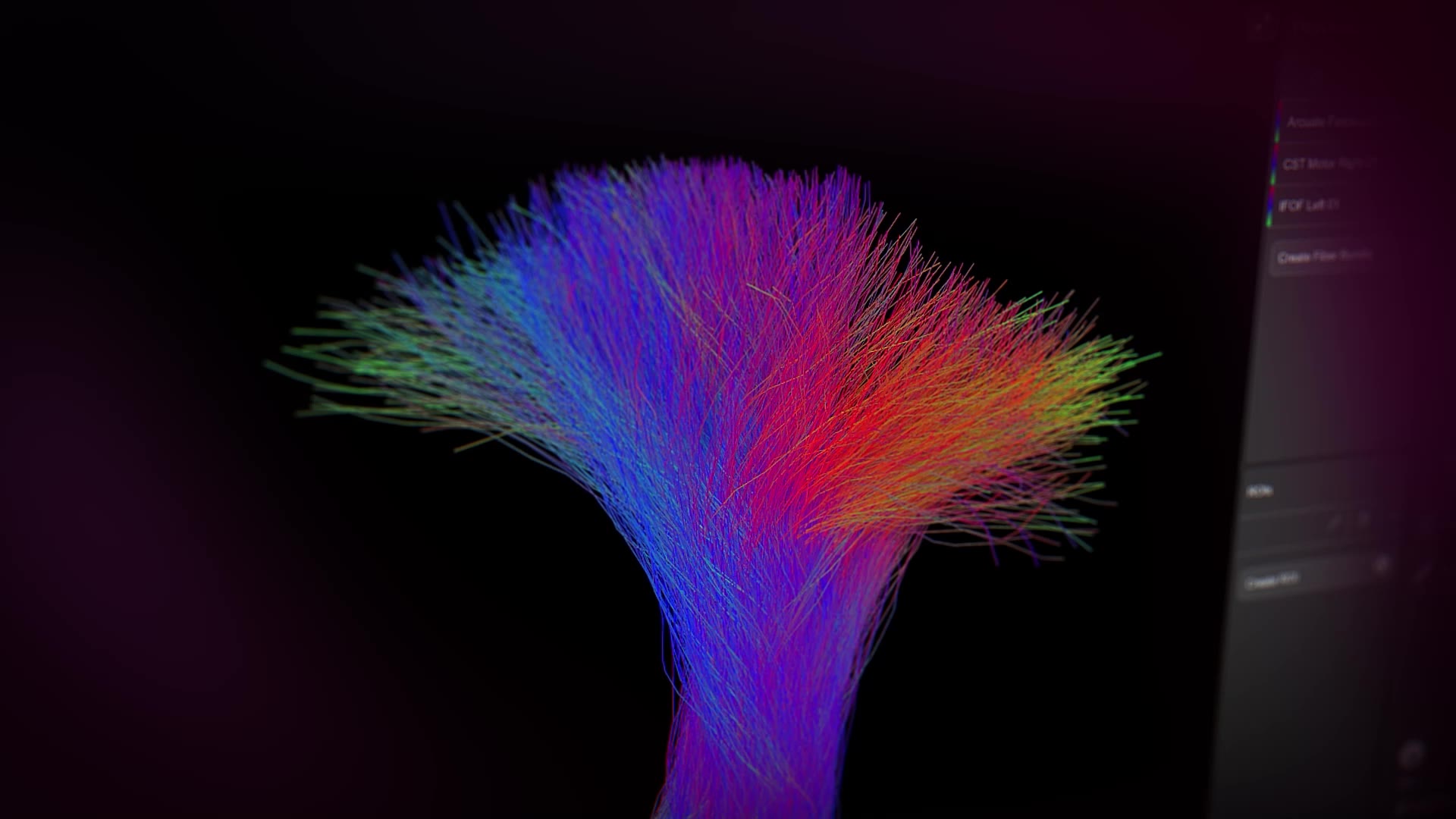
En plus du suivi déterministe des fibres, Elements Fibertracking vous offre la possibilité de voir encore plus avec l’ajout de la tractographie probabiliste des fibres. Basculez entre ces deux modes pour visualiser les faisceaux de fibres complexes à proximité des régions tumorales ou des œdèmes.1

Tractographie déterministe basée sur le DTI
Le suivi déterministe des fibres basé sur le DTI fournit un modèle detenseur simple et double, robuste, capable de reconstruire des voies simples, facilitant la visualisation et l’interprétation des résultats.
Tractographie probabiliste basée sur la CSD1
Le suivi probabiliste des fibres basé sur la CSD est une méthode de suivi avancée qui permet l’identification de plusieurs directions de fibre dans un voxel, surmontant la limitation de la technique basée sur le DTI lors de la traversée de fibres, de fibres en éventail ou fortement angulées, ou lors de la navigation dans des régions d’œdème.
Cas d’utilisation pour le suivi des fibres
Elements Fibertracking vous fournit les informations dont vous avez besoin pour prendre vos décisions éclairées.


Votre découverte des méandres du cerveau commence ici
Expérimentez les solutions chirurgicales de pointe.

Planification crânienne
Nos modules logiciels Brainlab Elements sont conçus pour aider les équipes cliniques dans la planification de pointe pour la chirurgie crânienne guidée par l’image.

Microscope Navigation
Microscope Navigation injecte des données pertinentes directement dans l’oculaire, fournissant aux chirurgiens un contexte significatif sans distraire de la situation chirurgicale.

Navigation crânienne
Notre système de navigation crânienne allie facilité d’utilisation et fonctionnalités étendues, permettant une planification éclairée de l’approche et une navigation fiable, adaptées aux besoins des chirurgiens lors des interventions crâniennes.
Pas encore commercialisé dans certains pays. Merci de contacter votre représentant des ventes.