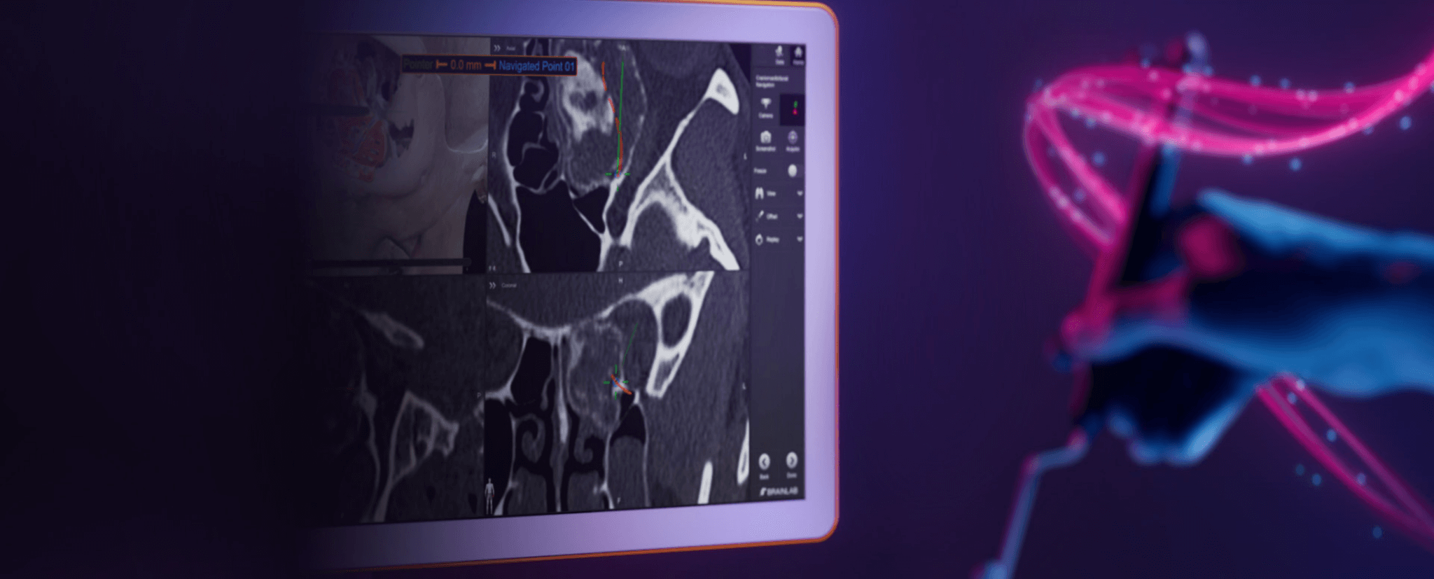
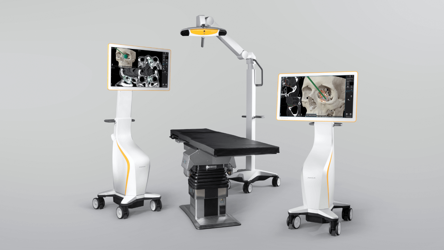
Header Image: Brainlab Craniomaxillofacial (CMF) Navigation

Craniomaxillofacial Navigation1Precision Guidance with Real-time Verification

Real-time realization of your surgical plan

Brainlab Craniomaxillofacial (CMF) Navigation enables you to achieve your desired outcomes for mid-facial reconstructions and tumor resections using real-time image-guided realization of the surgical plan. Clinicians can verify tumor removal, repositioned bone fragment and final implant positions. Intraoperative navigation and imaging allow you to stay on track throughout the procedure and be sure to avoid critical structures like the optic nerve.

How does our Craniomaxillofacial Navigation technology
help keep procedures on track?

Intraoperative plan realization

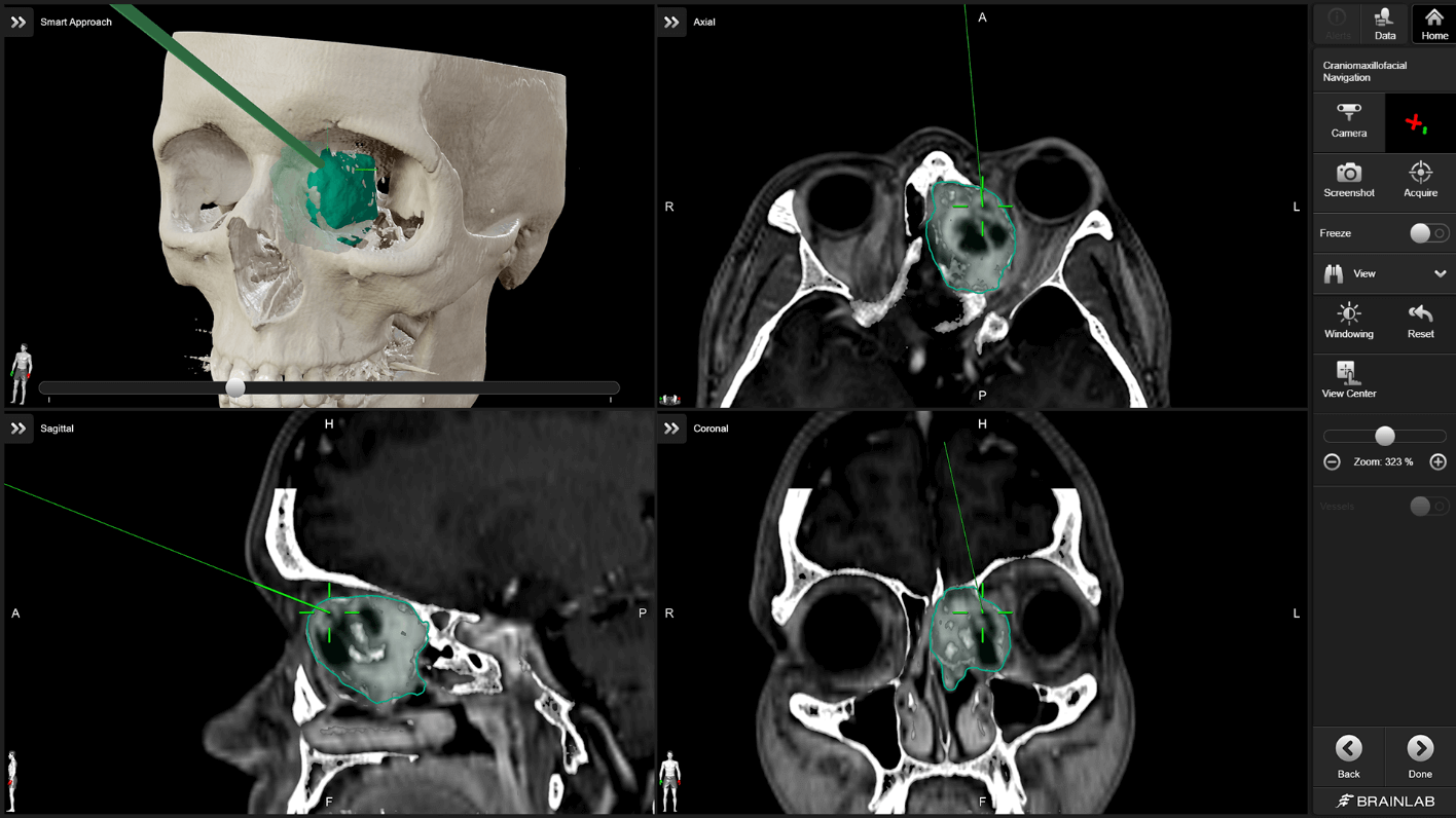
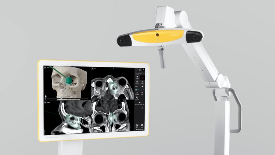
All relevant data planned with Elements software can be automatically transferred to the navigation system and utilized during surgery. The software provides a full feature set for indications like tumor resections or mid-facial reconstructions and offers accurate information about the position of fragments, mesh and implants.

Screenshot: Intraoperative Plan Realization with Elements Software

Implant position verification with Replay

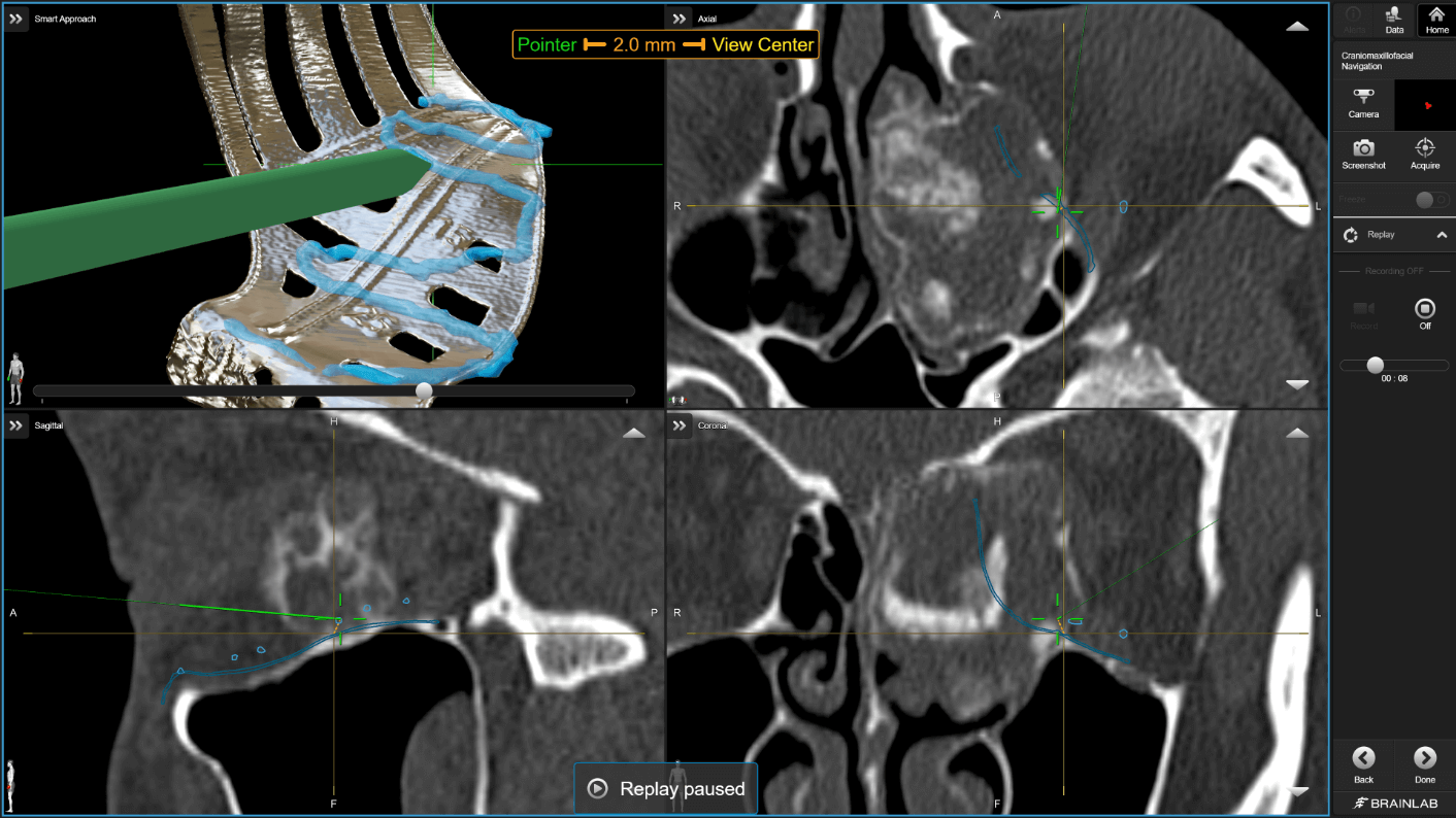
Compare planned to actual position of implants, mesh and bone fragments. With Replay navigation pointer functionality, you can intraoperatively trace and verify surfaces and landmarks to optimize positions of any biocompatible materials, all prior to fixation.

Screenshot: Replay navigation pointer functionality

Individually configured views

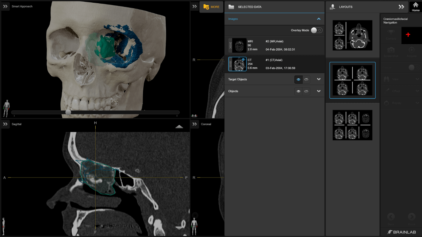
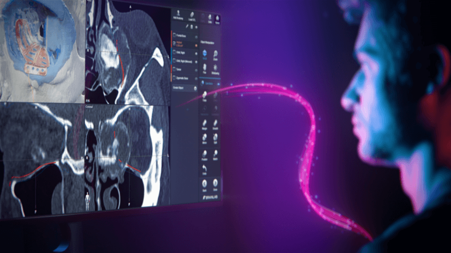
Drag-and-drop functionality allows you to choose from different layout options and configure your display to best view your patient data, including CT, MR and dynamic 3D. Peel through different layers—from skin and bone to pre-planned and target structures like tumor and implants.

Screenshot: Brainlab Craniomaxillofacial (CMF) Navigation - Individually configured views

Intraoperative results control

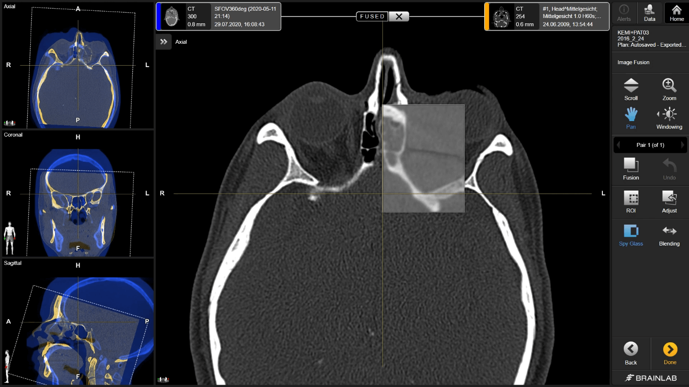
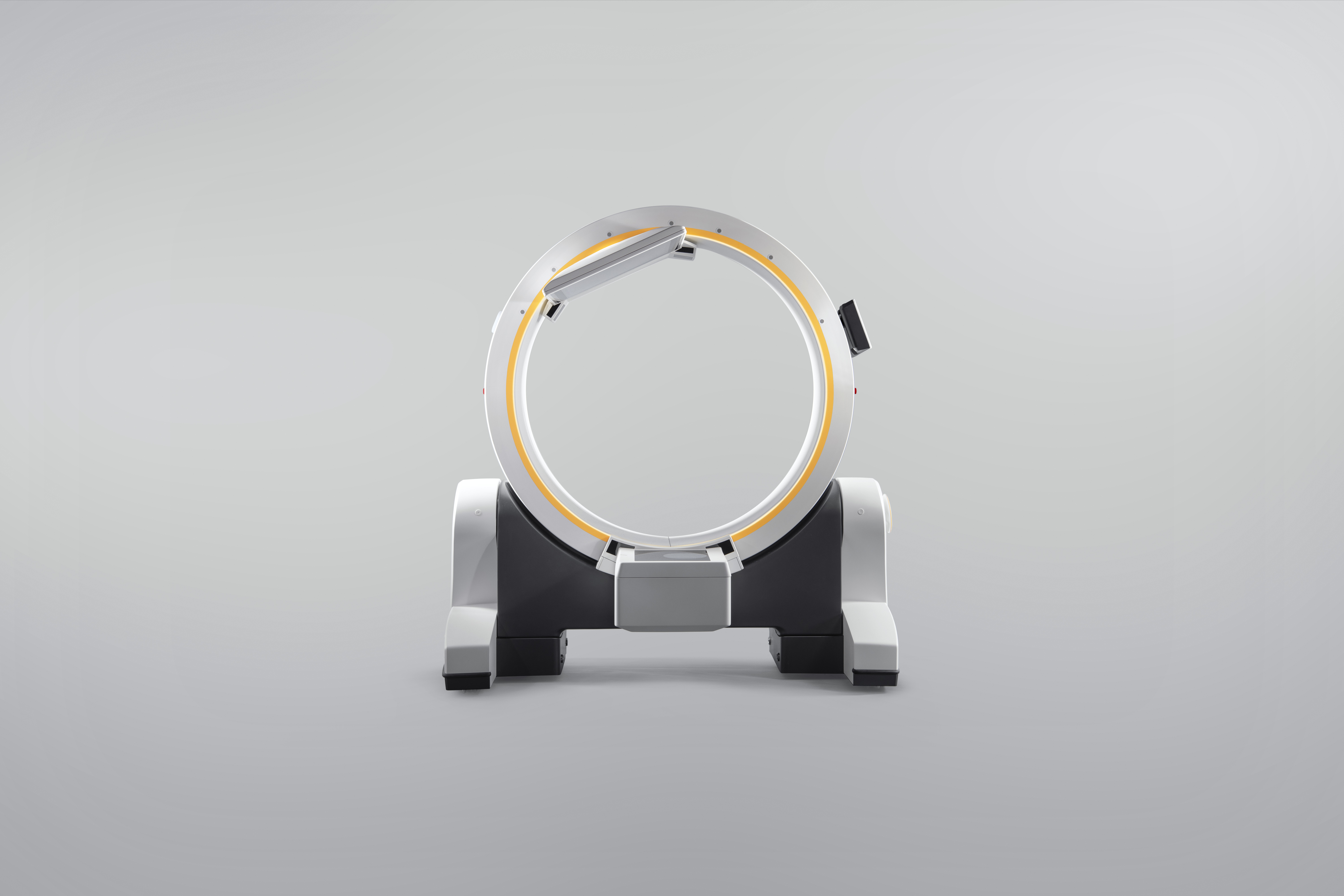
For final quality control in the O.R., CMF Navigation can be combined with imaging systems, like our Loop-X Mobile Imaging Robot and other third-party devices. Intraoperatively acquired scans can be automatically loaded onto the navigation system and fused with your preoperative plan for instant results verification.

Screenshot: Intraoperative Results Control with CMF Navigation and Loop-X Mobile Imaging Robot

Improved functional outcomes and aesthetics

Craniomaxillofacial Navigation supports clinicians in localizing critical structures, like the optic nerve or posterior ledge. It improves functional outcomes by reducing diplopia or enophthalmos and restores facial symmetry beyond conventional surgical techniques.

  • Transfer all planned data with Brainlab Elements to the navigation system for image-guided realization

  • Avoid critical structures and arrive at target structures with enhanced visualization and real-time instrument tracking

  • Improve functional outcome and aesthetics compared to conventional surgeries with navigation guidance along the reconstruction

Are you ready for real-time realization of your CMF surgical plan?

Flexibility with Craniomaxillofacial Navigation includes a broad range of registration options and integration of imaging devices and instruments to fit your procedure requirements.

Surface matching

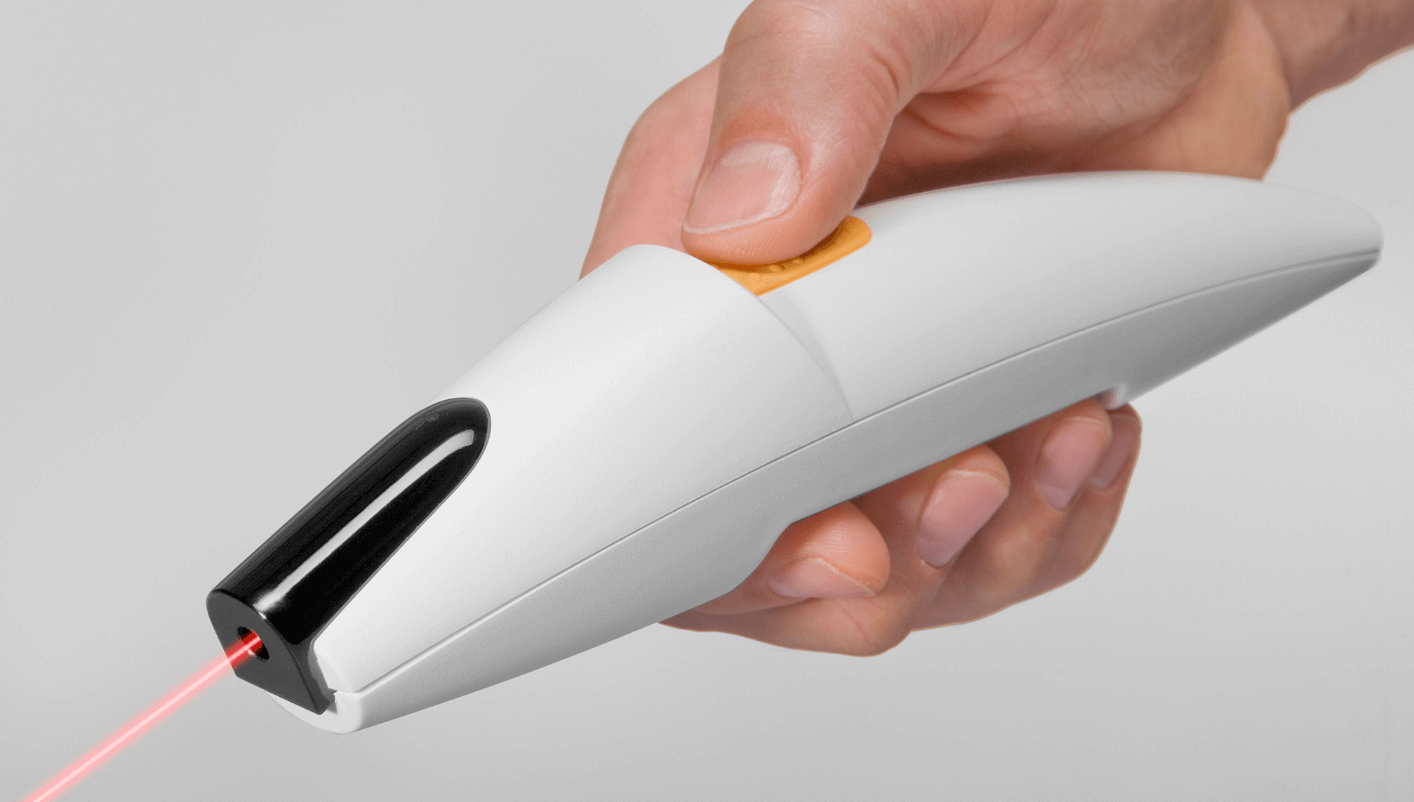
Surface matching is a fast registration method based on a preoperative CT or MR scan. Registration is performed by simply scanning the surface of the patient’s head using Z-touch®, a contact-free laser pointer, or Softouch®, a touch-sensitive pointer. An auto-correcting algorithm matches the acquired points with the preop scan in just a few seconds.

Z-touch®, a contact-free laser pointer

Landmark registration

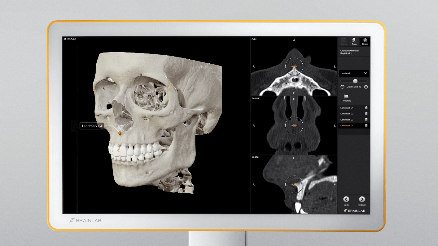
Landmark registration uses a paired point matching technique to reference your patient’s anatomy to the loaded data. Registration points can both include anatomical landmarks or fiducial markers, using screws or dental splints. After defining the registration points in the patient’s image set, the navigation pointer is used to acquire the points on the patient’s anatomy thereby correlating the corresponding image data.

Screenshot: Landmark Registration - Paired Point Matching Technique for Patient Anatomy Referencing

Automatic integration of intraoperative images

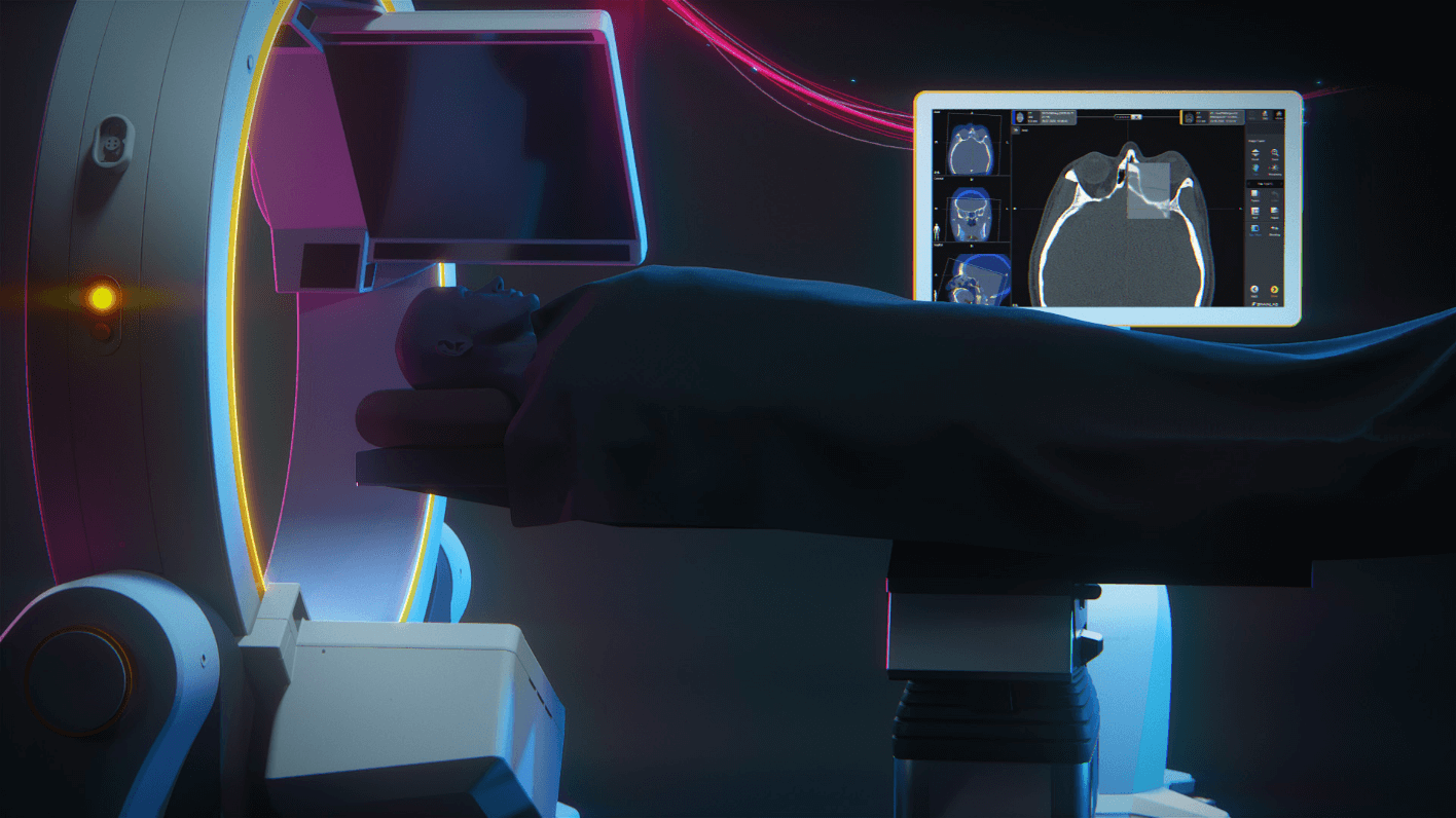
Automatic Image Registration (AIR) allows for the integration of intraoperative CT and cone-beam CT images with Craniomaxillofacial Navigation. Intraoperative CT scanners, like Loop-X Mobile Imaging Robot, can be integrated with reflective markers to enable tracking by the navigation system. In addition, the isocenter of the scanner is calibrated to provide automatic registration of the images upon delivery of the scans to the navigation system. AIR optimizes your workflow by providing faster registration compared to manual registration methods.

Illustration: Loop-X Mobile Imaging Robot for Automatic Intraoperative Image Integration with Craniomaxillofacial Navigation

What our customers are saying2

How Brainlab technologies support the craniomaxillofacial workflow

During complex TMJ reconstructions, navigation helps for orientation, dissection and drilling – so you know where you are in the lateral skull base, especially in abnormal anatomy. Additionally, if you have the STL files of your TMJ prosthesis, you can play the depth of your dissection, the drilling vectors and the extent of resection towards the medical skull base so that your implant immediately fits. This saves surgical time and is easy on your nerves.

Rüdiger Zimmerer, Priv.-Doz. Dr. Dr., University Medical Center Leipzig, Germany
Photo of Priv.-Doz. Dr. Dr. Rüdiger Zimmerer, University Medical Center Leipzig, Germany

Brainlab navigation enables us to realize preoperative plans. All relevant data is immediately available and can be utilized during surgery. This allows us to perform reconstructive procedures with less radiation for high precision verification of the final implant position.

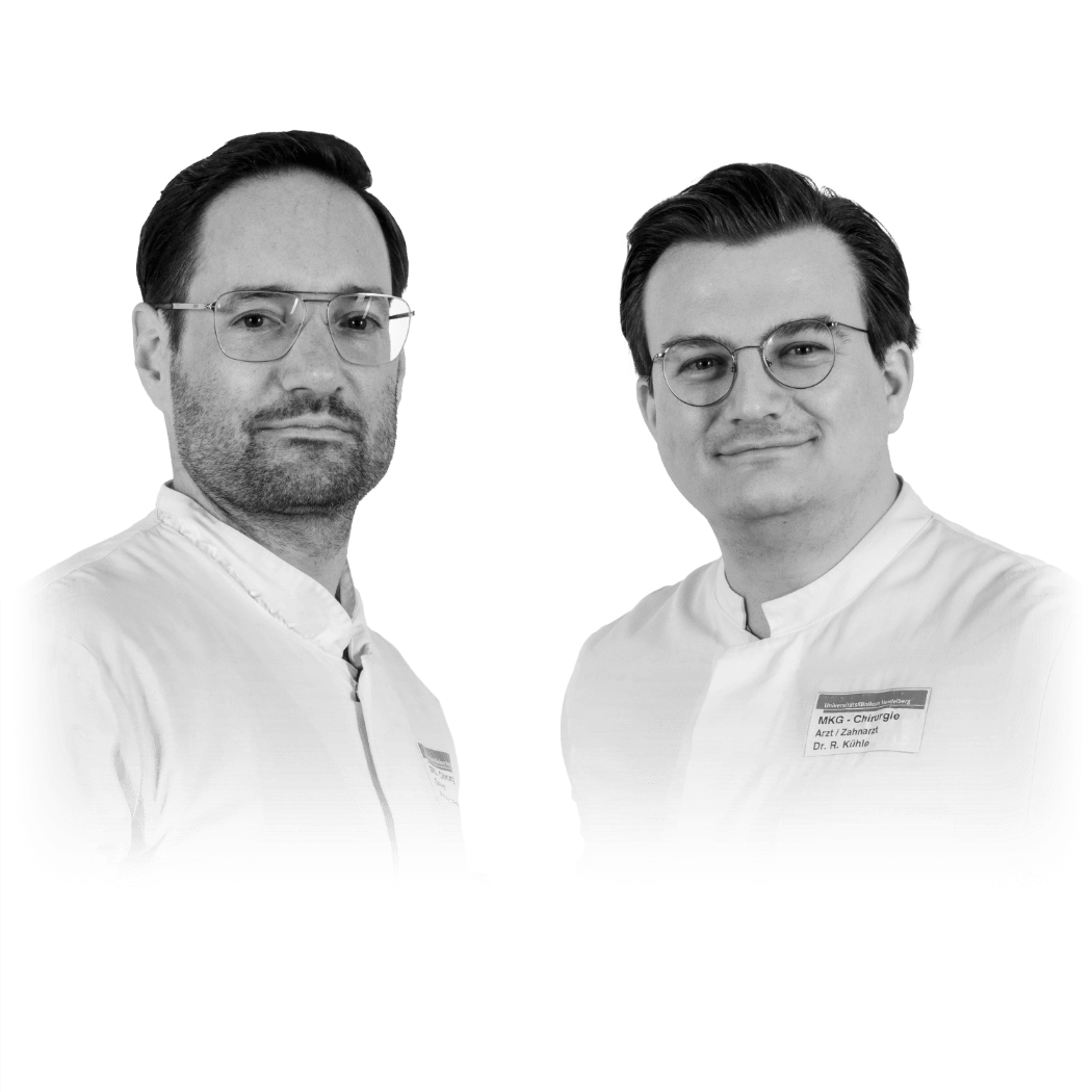
Prof. Christian Freudlsperger, MD, DDS & Reinald Kühle, MD, DDS, Heidelberg University Hospital, Germany
Photo of Prof. Christian Freudlsperger, MD, DDS & Reinald Kühle, MD, DDS, Heidelberg University Hospital, Germany

Craniomaxillofacial planning software improves my daily work especially in complex mid-facial reconstructions and tumor treatments. Additionally, the transfer of the planned data to intraoperative systems like imaging or navigation platforms increases my clinical outcome as I can perform a live intraoperative check of my preoperative plan.

Professor Alexander Schramm, MD, DDS, University and Military Hospital Ulm, Germany
Photo of Professor Alexander Schramm, MD, DDS, University and Military Hospital Ulm, Germany

The combination of surgical navigation and intraoperative imaging with Loop-X gives me constant control of my outcomes. Immediate progress verification in the O.R. can even prevent complications and revision surgeries.

Simon Enzinger, MD, DMD, University Hospital Salzburg, Austria
Photo of Simon Enzinger, MD, DMD, University Hospital Salzburg, Austria

During complex TMJ reconstructions, navigation helps for orientation, dissection and drilling – so you know where you are in the lateral skull base, especially in abnormal anatomy. Additionally, if you have the STL files of your TMJ prosthesis, you can play the depth of your dissection, the drilling vectors and the extent of resection towards the medical skull base so that your implant immediately fits. This saves surgical time and is easy on your nerves.

Rüdiger Zimmerer, Priv.-Doz. Dr. Dr., University Medical Center Leipzig, Germany
Photo of Priv.-Doz. Dr. Dr. Rüdiger Zimmerer, University Medical Center Leipzig, Germany

Brainlab navigation enables us to realize preoperative plans. All relevant data is immediately available and can be utilized during surgery. This allows us to perform reconstructive procedures with less radiation for high precision verification of the final implant position.

Prof. Christian Freudlsperger, MD, DDS & Reinald Kühle, MD, DDS, Heidelberg University Hospital, Germany
Photo of Prof. Christian Freudlsperger, MD, DDS & Reinald Kühle, MD, DDS, Heidelberg University Hospital, Germany

Flexible technologies to complement your CMF surgeries

Close-up image of Curve Navigation

Curve Navigation

Go beyond image guidance for effortless insights

Loop-X

Experience new surgical possibilities

Header Image: Brainlab Craniomaxillofacial Planning software

Craniomaxillofacial Planning

Customize planning with Brainlab Elements

Discover our clinical studies and on-demand CMF webinars on the benefits of navigation

Clinical literature library

Advancing medical technology through scientific evidence

Brainlab Clinical Affairs conducts advanced research and gathers scientific evidence to help create safe, efficient, cost-effective medical technology.

Craniomaxillofacial Webinar Series

Explore our collection of on-demand CMF webinars

Presented by leading clinicians in the field of craniomaxillofacial surgery, our webinars discuss the advantages of navigation in CMF procedures like TMJ reconstructions, tumor surgeries and more.
  1. Not yet commercially available in several countries (CE mark available). Please contact your sales representative

  2. The statements of the surgeons reflect only personal opinions and experiences and do not necessarily reflect the opinions of Brainlab or any affiliated institution.