An individual using the Mixed Reality Viewer in an office, examining a cranial image

Mixed Reality Viewer for NeurosurgeryView brain anatomy
at every angle

Discover more

Dive into the details of neuroanatomy. The Mixed Reality Viewer1 is a technology that works in tandem with Brainlab Elements to produce hyper-realistic 3D patient data. Easy to integrate into surgical planning workflows, this software offers a fully immersive environment for surgeons to understand, visualize and prepare for neurosurgical cases.

1

Enhanced surgical planningreview with patient-specific 3D data

The Mixed Reality Viewer gathers all relevant medical images and patient-specific anatomical structures to produce accurate and detailed 3D models. With the 3D anatomy displayed through the Magic Leap 2 device, surgeons can prepare for procedures by examining, reviewing and interacting with the data.

2

Increased access to immersivelearning

Hands-on, collaborative and accessible medical education starts with the Mixed Reality Viewer. Both residents and medical students can benefit from the immersive 3D environment and gain deeper insights into patient anatomy.

3

Individual patient treatmentplans move from screen to reality

Brain operations are highly complex procedures. The Mixed Reality Viewer can thereby be used to answer patient questions by clearly depicting individual diagnoses and surgical treatment plans. 

workflow_background.jpg

Experience our vision for mixed reality

Explore the features of mixed reality for neurosurgery

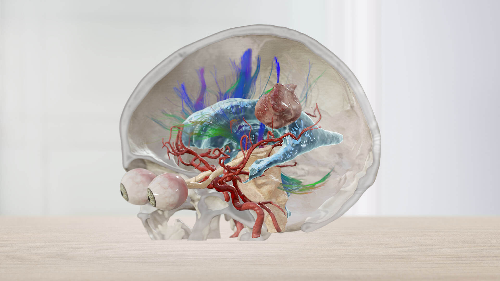
When it comes to neurosurgical procedures, mixed reality technology enables healthcare professionals to gain a better understanding of a patient’s brain—layer by layer. This includes gaining insights into the location of a deep-seated tumor and identifying critical surrounding anatomical structures like the optical nerve, brainstem and fiber tracts.

Mixed Reality Viewer for neuro-oncology

  • Position the 3D patient model at the preferred operating angle, simulate the craniotomy and review the planned trajectory to the tumor.

  • Overlay and scroll through 2D data on the 3D model along the trajectory path to view the structures at risk from any angle, providing awareness of the anatomical spatial surroundings.

  • Increase understanding of functional anatomy and vascular structures around the tumor and their displacement in relation to the planned approach before entering the O.R.

  • Benefit from having the same 3D model available on a navigation system intraoperatively.

03_cranial.jpg

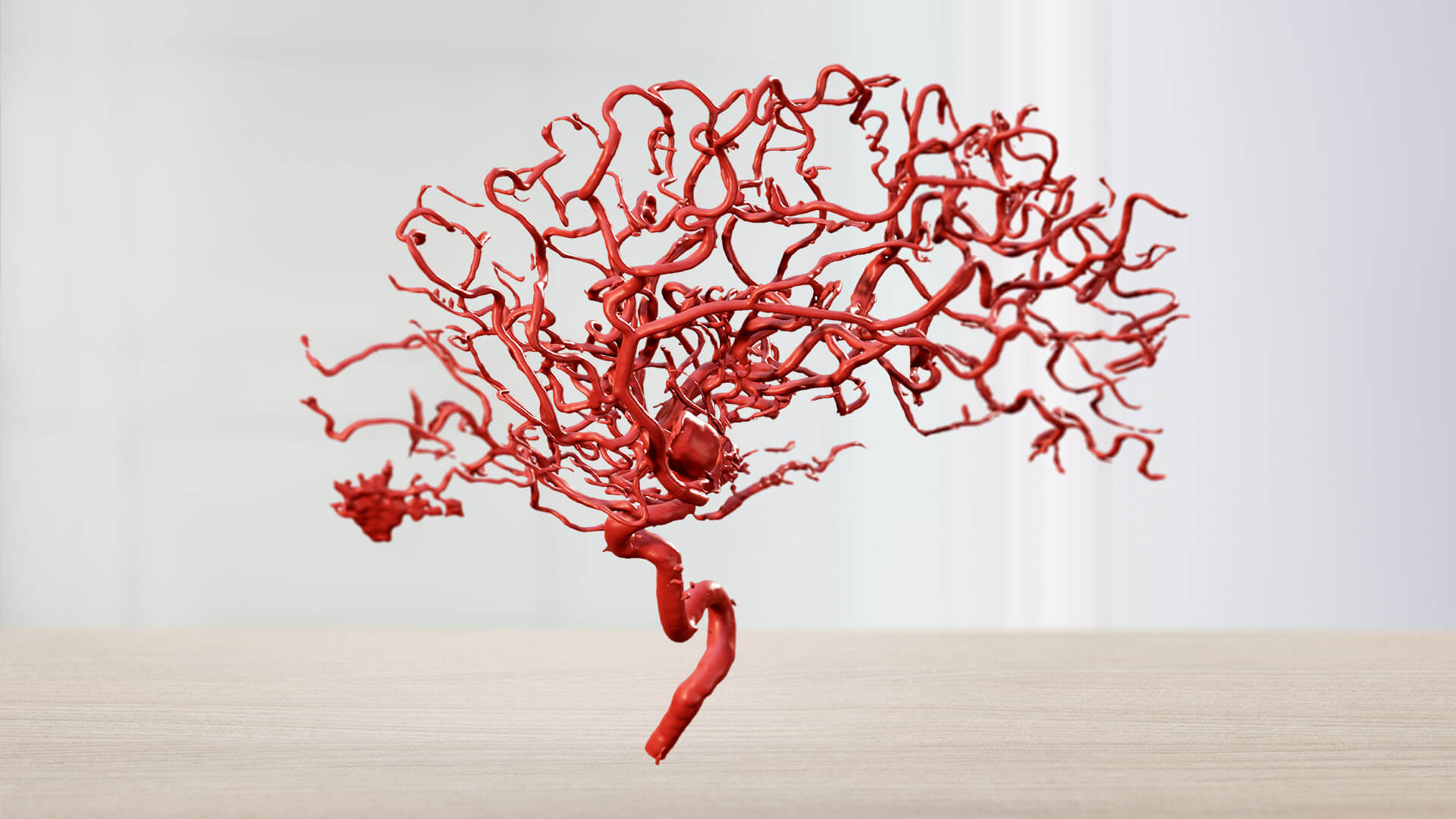
Mixed Reality Viewer for vascular neurosurgery

  • Combine the power of Elements Image Fusion Angio, Elements SmartBrush Angio and the Mixed Reality Viewer to review a patient’s vasculature.

  • Outline the size of a craniotomy, transfer points and measurements during planning review from the Mixed Reality Viewer to the patient data for intraoperative use.

  • Anticipate the size and positioning of a vascular clip before entering the O.R. by understanding the pathology through hyperrealistic three-dimensional visualization.

Showcasing the Mixed Reality Viewer for Vascular Indications

Find your future workflow today

Expand your perspective with the Mixed Reality Viewer

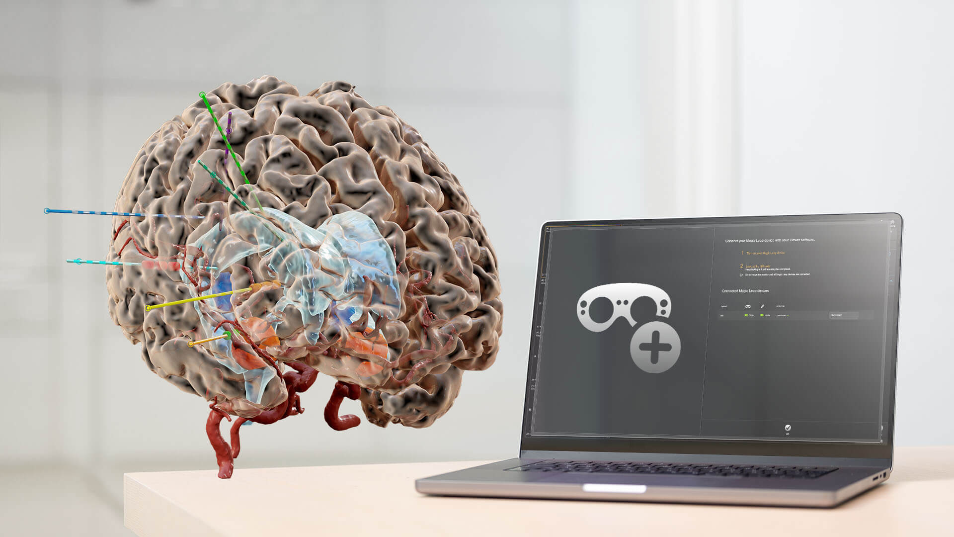
With a single click and a glance, your room is digitized for spatial computing. Images are transferred from the Elements Viewer software on screen into the room with the help of the Magic Leap spatial computing platform.

Start with a click

A single click on the Elements Viewer software opens up the world of mixed reality and a new, immersive way to understand patient data.

Elements Viewer software on a laptop screen

Customizable hardware features

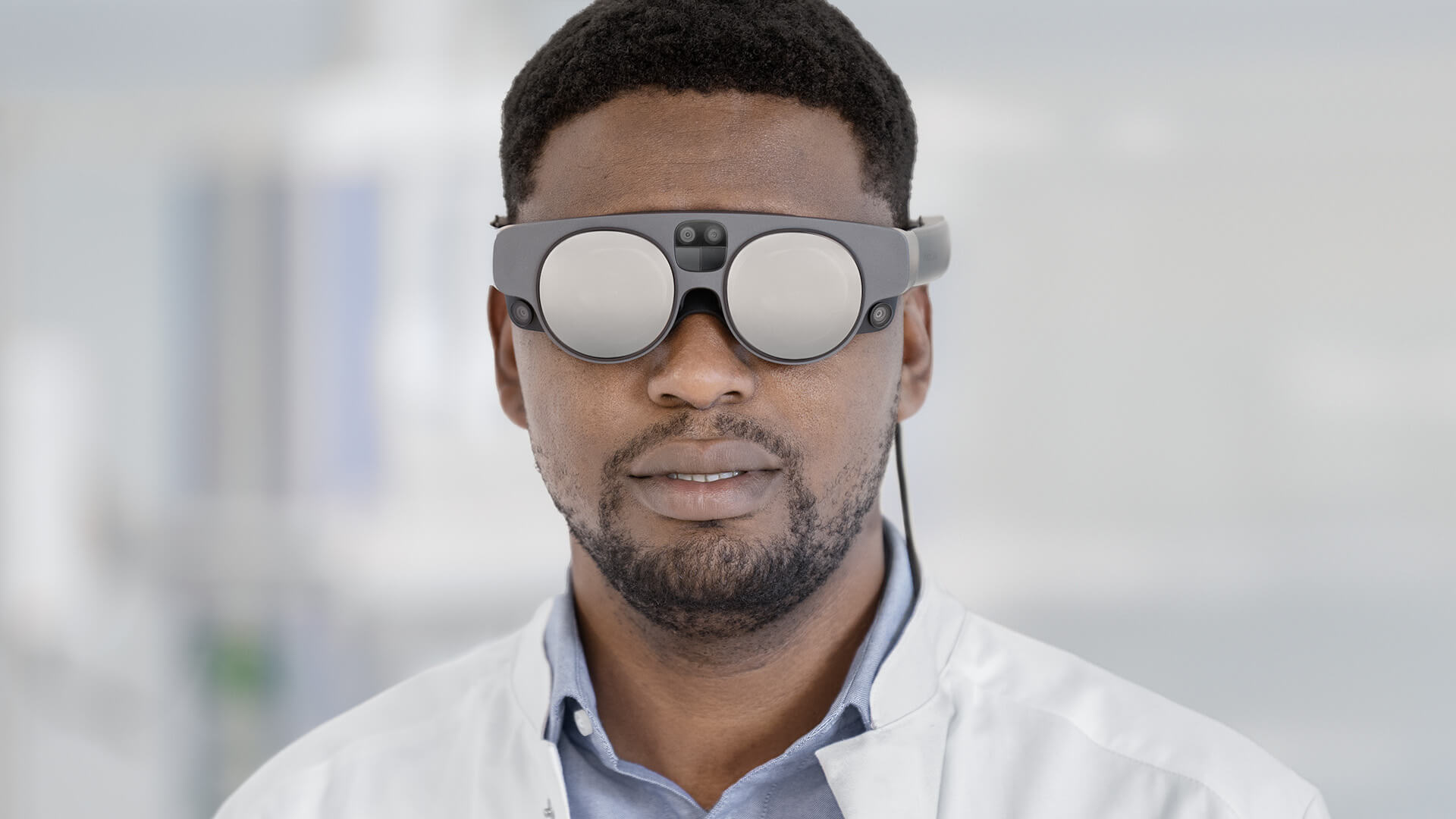
Cameras in the Magic Leap 2 scan and map the room, allowing 3D models and 2D slices to be displayed according to the surgeon’s preference.

Person wearing Magic Leap 2 - Mixed Reality Viewer by Brainlab

Assess, interact, learn

The virtual pointer makes it easy to interact with and highlight the image data and anatomical structures.

Virtual Pointer in use with the Mixed Reality Viewer, highlighting image data and anatomical structures
workflow_background.jpg

Easy integration into unique
clinical workflows

Illustration: a visual representation of brain anatomy and an arrow indicating how to start the Elements Viewer

1. Start Elements Viewer

Access patient data from PACS and load it into the Elements Viewer

Illustration of the Magic Leap 2 headset

2. Boot your Magic Leap Device

Boot your Magic Leap 2 headset and connect it to WiFi

Illustration of a laptop with a QR code on the screen

3. Scan QR Code

Scan the QR code on the screen with your headset

Illustration: a visual representation of brain anatomy and a controller indicating to step into an immersive 3D model using the Mixed Reality Viewer

4. Step into an immersive 3D model

Dive into patient data and interact with both virtual 2D images and 3D models

Brainlab's Mixed Reality Viewer in action, utilized by two individuals in an office setting

Mixed Reality Viewer PlatformVisualize Patient Data
in 3D

Read scientific literature about mixed reality

Teaser Image - Study - MRgFUS Planning with Elements Fibertracking

Clinical Paper

Usability of Mixed Reality in Awake Craniotomy Planning

Teaser Image - Study - Automatic Segmentation of the STN for DBS

Clinical Paper

Application of Augmented Reality in Trigeminus Neuralgia

Teaser Image - Clinical Literature Library

Clinical Paper

Virtual dissection of white matter pathways

  1. Not yet commercially available in several countries. Please contact your sales representative.

Sign up now, see the future of medtech later

Our innovative surgical solutions are always just one click away