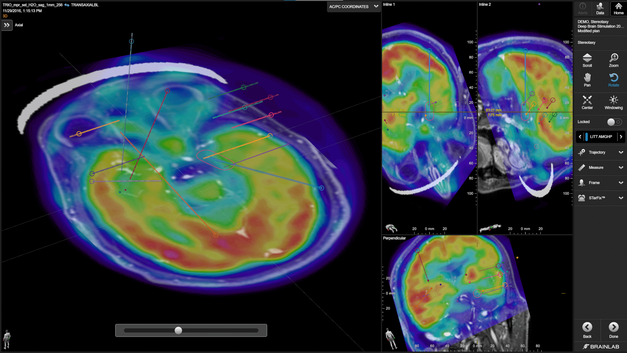
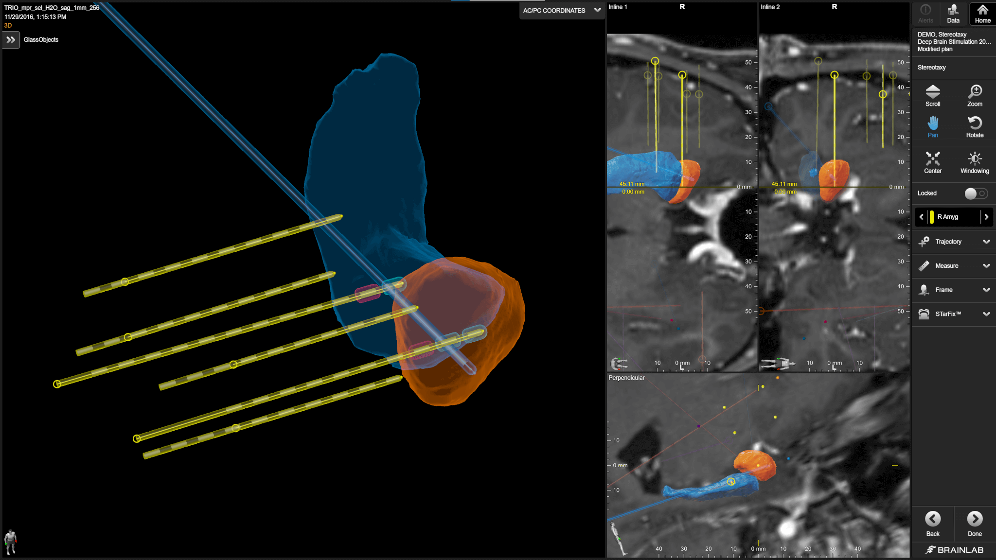
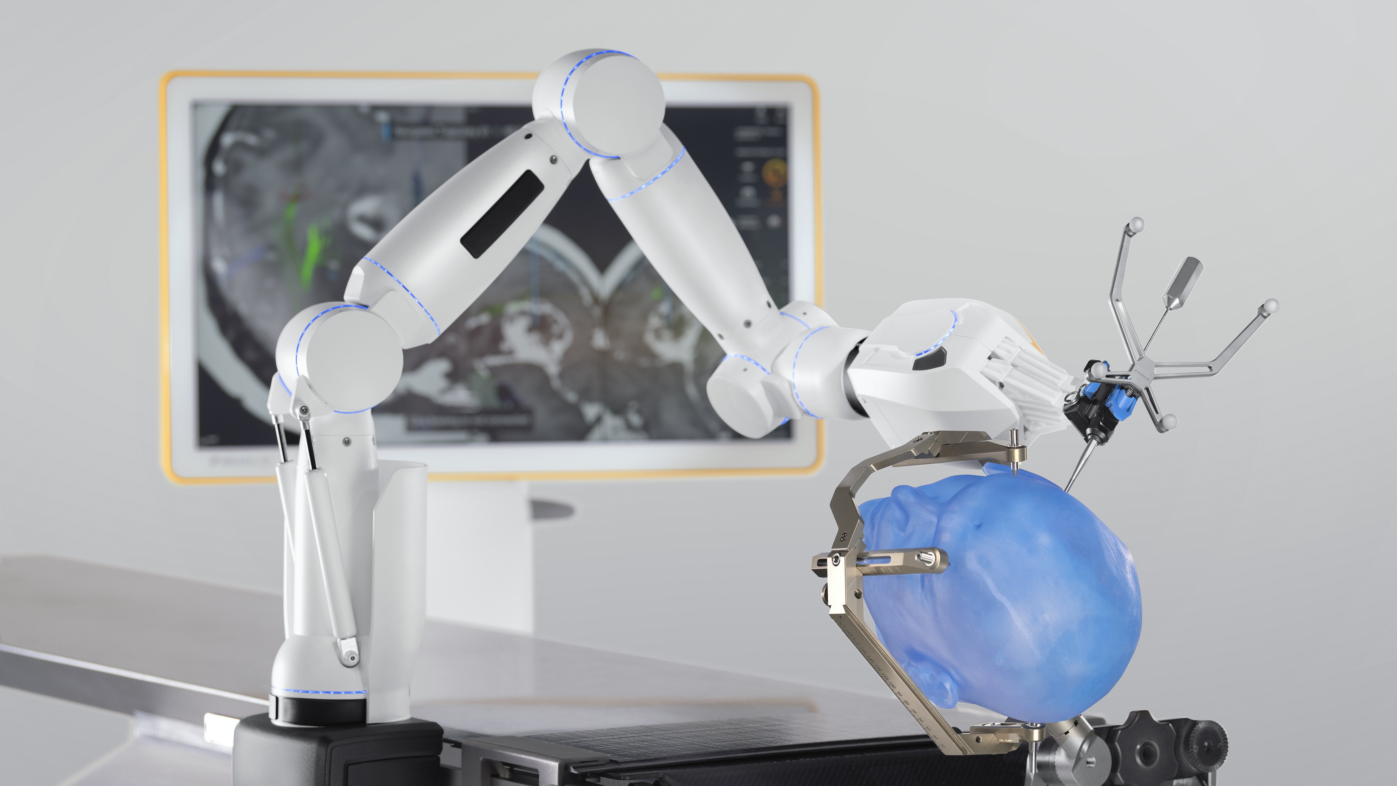
Brainlab epilepsy solutions bridge the gaps between hypothesis formulation, advanced sEEG planning, electrophysiology information and the navigation of surgical platforms for streamlined treatment execution and communication between clinical teams.

3D visualization is important for [epilepsy] resection surgery. I utilize Brainlab automatic segmentation to help me choose the surgical approach based on anatomical variations and to help orient myself.
Bring the best into surgery.
Bring Brainlab.
Contact us today to experience state-of-the-art solutions for surgery.
Managing epilepsy –
from supporting diagnosis to surgical intervention and postop review
Phase I – Preparation for sEEG
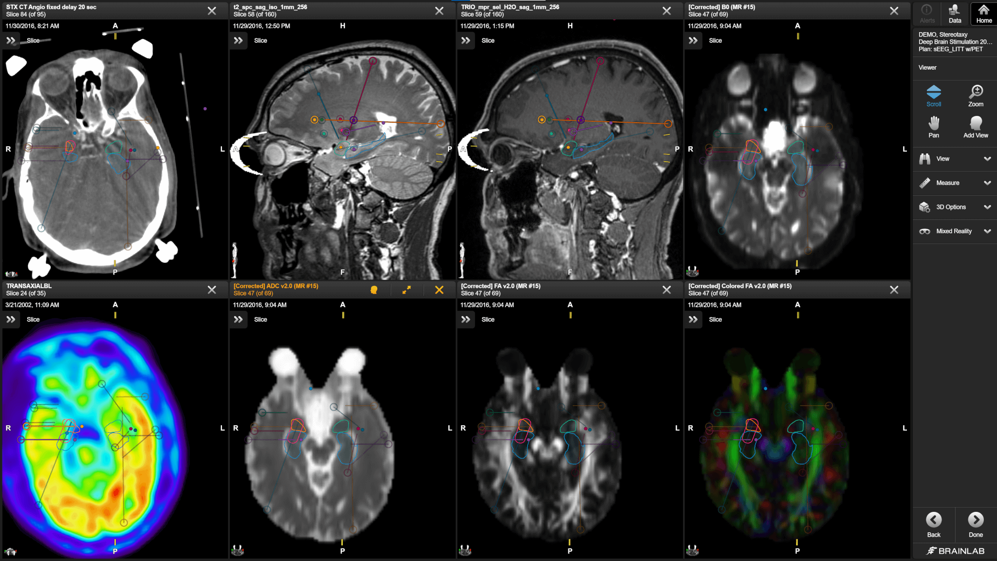
Review & share all patientimaging & information
The DICOM Viewer supports multi-modal diagnostic imaging and provides a single environment to review and share all relevant patient imaging for hypothesis formulation. Best-in-class image fusion software provides accurate co-registration to allow simultaneous review and seamless blending of datasets.
Visualize and target specific structures
Elements Object Manipulation enables users to visualize patient-specific structures created from an advanced voxel-by-voxel analysis of the patient’s imaging studies and bring these objects into trajectory planning.
sEEG planning made easy
Advanced trajectory planning and modeling tools facilitate the visualization of the exact location of planned sEEG leads and the precise location of the recording contacts within patient anatomy using user-built 3D electrode models.
Phase II – sEEG implantation surgery
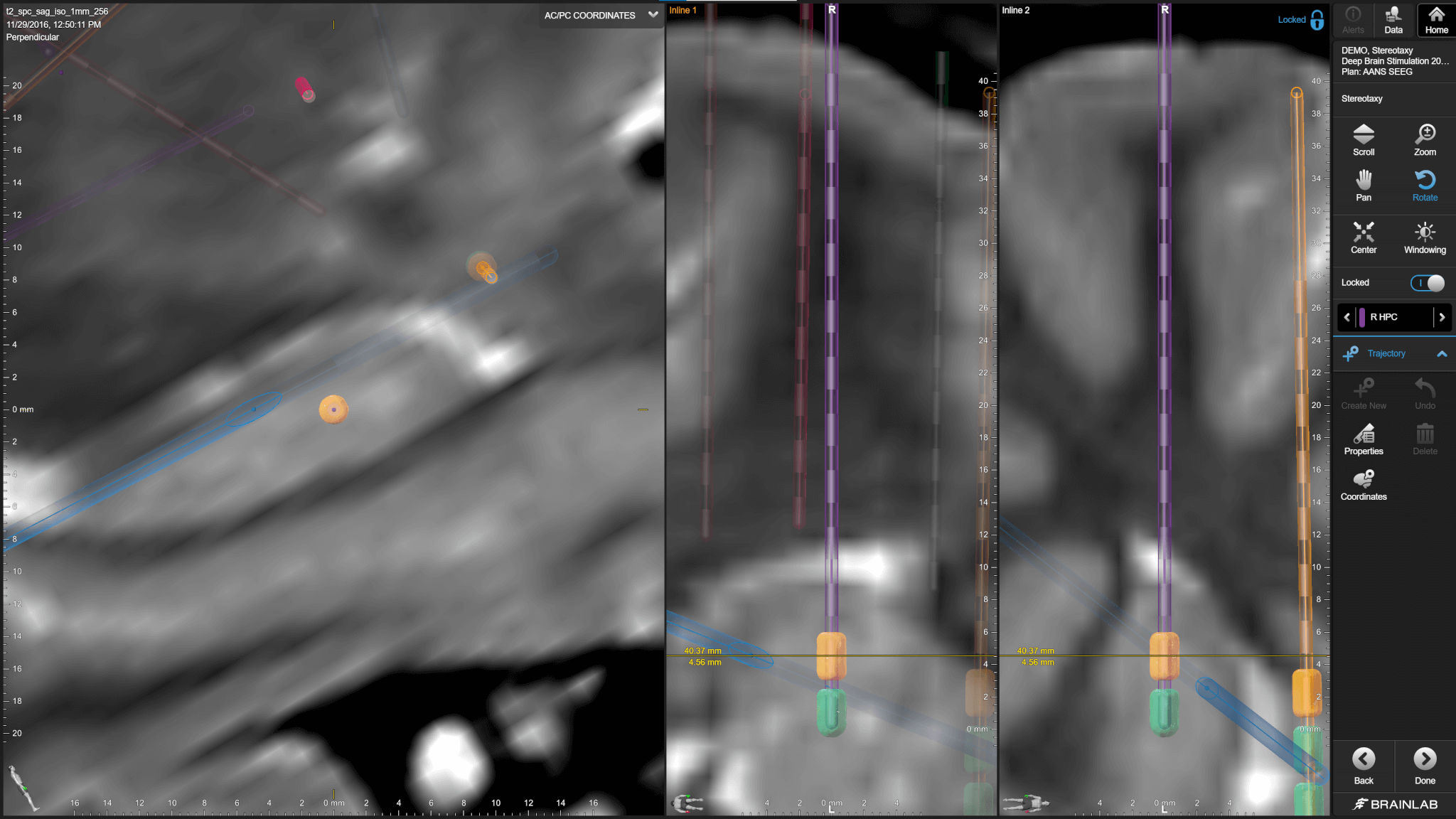
Support from planning to surgical implantation
With Brainlab Alignment Software Cranial and the Cirq robotic platform, users can seamlessly bring sEEG trajectory workflows from planning to surgical implantation through integrated hardware and software solutions.
Automatic intraoperative imaging capabilities
Either individually or together as part of the Brainlab Functional Neurosurgery Robotic Suite, users can leverage the powerful intraoperative imaging capabilities of the Loop-X® robotic imaging device for patient registration in the O.R. through Automatic Image Registration (AIR), frame localization or bone fiducial visualization to save time and enhance accuracy.
Rapidly and reliably implant sEEG leads
When using the Cirq Arm System, take advantage of a modular, rail-mounted robotic system that utilizes proven optical navigation technology to reliably implant sEEG leads under live tracking to ensure accuracy at every step.
Phase III – Turning sEEG results into surgical treatment planning
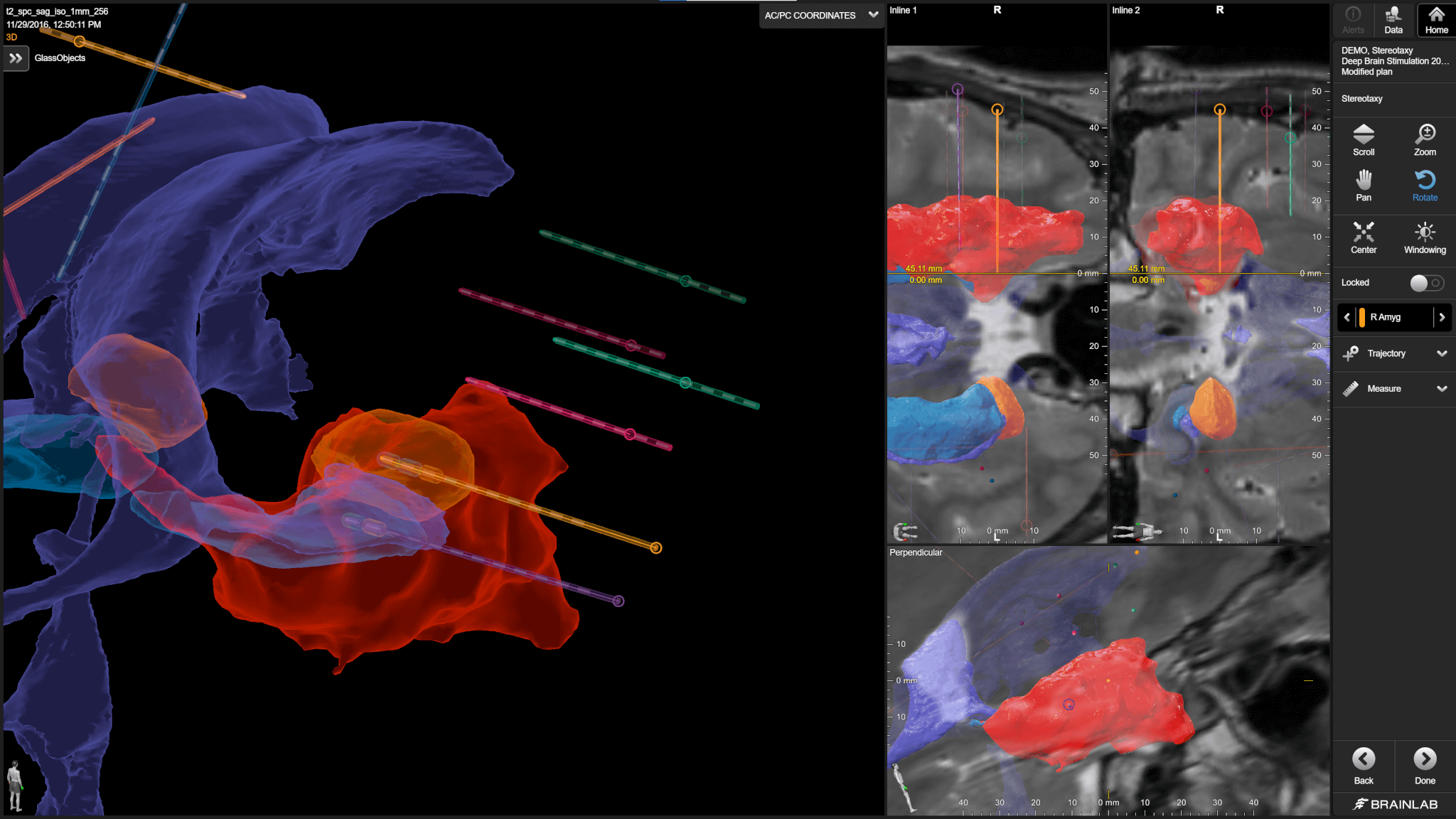
Automatically localize implanted sEEG electrodes
Following sEEG implantation, automatically verify the placement of trajectories with Elements Lead Localization and determine individual contact locations on the leads via powerful 3D visualization.
Interact and collaborate on your surgical plan in a brand-new way
With the Mixed Reality Viewer, unlock a powerful tool for collaboration in preparation for surgical planning. Use 3D visualization of specific patient structures to aid in reviewing the surgical approach and orientation to specific patient anatomical variations.
Seamless integration from planning to surgical intervention
Bring your plan full circle with advanced Brainlab cranial navigation solutions that enable the most comprehensive image guided surgical treatment, utilizing microscope integration, ultrasound navigation, imaging and more.
Innovation isn’t often free, but our demos are.
Find your future workflow today.
Cirq in combination with the Alignment Software Cranial for sEEG procedures is not yet commercially available in several countries. Please contact your sales representative.
